
Research Article Summary
5 Minute Read
WUT researchers build a vascularized ovarian-cancer-on-a-chip using 3D printed resin molds
Title
An Advanced 3D Model of Vascularized Epithelial Ovarian Cancer in a Tumor-on-a-Chip System Based on Multi-Cell Culture
Authors
Magdalena Flont, Agnieszka Żuchowska, Oliwia Tadko, et al.
Journal
Sensors (MDPI), 2026, Vol. 26, Article 1503
DOI Link
Summary
Researchers at the Warsaw University of Technology fabricated a multilayer tumor-on-a-chip that recreates a 3D, vascularized model of epithelial ovarian cancer, using 3D-printed resin molds to cast the PDMS device. Flont et al. show that endothelial cells will grow into a hollow vessel and migrate toward the tumor layer, reproducing early angiogenesis on-chip.
Summary Author
This page was prepared by CADworks3D to summarize and highlight a peer-reviewed research article authored by independent researchers utilizing the CADworks3D system.
Title
An Advanced 3D Model of Vascularized Epithelial Ovarian Cancer in a Tumor-on-a-Chip System Based on Multi-Cell Culture
Authors
Magdalena Flont, Agnieszka Żuchowska, Oliwia Tadko, et al.
Journal
Sensors (MDPI), 2026, Vol. 26, Article 1503
DOI Link
Key Results at a Glance
50–56 µm
Membrane thickness
The thin PDMS layer separating the two cell layers
370 ± 9 µm
Vessel diameter
Close to a small vein in the body
> 200 µm
Cell migration
Distance endothelial cells traveled toward the tumor
10 days
Culture lifespan
Device stayed stable and functional throughout
Objective
Ovarian cancer is one of the deadliest cancers in women, largely because it is usually caught late and resists chemotherapy. A big reason new treatments are hard to test is that the standard lab tool, cells grown flat on a dish in two dimensions, does not behave like a real tumor. A real tumor is a 3D community: cancer cells, support cells called fibroblasts, blood vessels, and a surrounding scaffold of proteins, all signaling to each other. Flatten that into a single layer and you lose almost everything that makes a tumor act like a tumor.
Researchers have tried to close this gap with 3D spheroids and hydrogel cultures, and these are an improvement, but most of them still leave out two features that matter a great deal in ovarian cancer: a heterogeneous, layered architecture and a working blood supply. Tumors actively recruit new blood vessels by releasing signaling proteins such as VEGF, and that vessel growth (angiogenesis) feeds the tumor and opens routes for it to spread. A model that cannot reproduce vessel growth cannot realistically predict how a drug, especially an anti-angiogenic drug, will perform.
The gap this paper addresses is therefore a build problem as much as a biology problem. The team set out to construct a single microfluidic device that holds three separate but connected environments at once: a channel for cancer cells, a channel for fibroblasts, and a channel where a real hollow blood vessel can form. Crucially, the two cell layers had to be separated by a membrane thin enough to act like the basement membrane found under real epithelial tissue, while still letting the two sides communicate. Hitting those geometric targets, channels on the order of hundreds of µm with a membrane only tens of µm thick, is what pushed the team toward a precise, repeatable molding process rather than hand assembly.
Methodology and Design
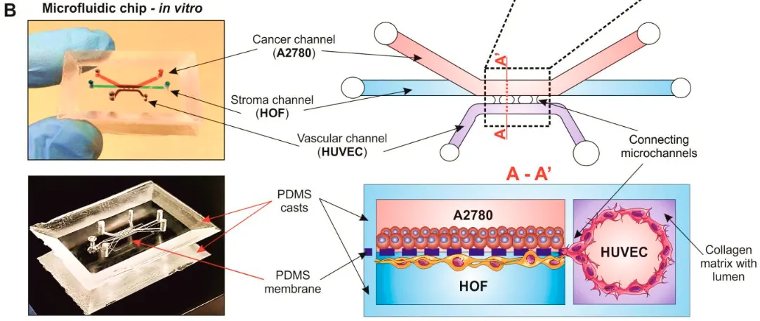
The device is a two-layer PDMS chip separated by a thin PDMS membrane, and the whole thing starts from a mold. The team designed the channel network as a CAD model, then 3D-printed the mold in a resin specifically formulated for casting silicone. Liquid PDMS was poured into the printed mold, cured, and peeled out as a finished cast.
That mold became the foundation for four distinct functional zones built into the chip:
Cancer Channel
Stromal Channel
Vascular Channel
Connecting Microchannels
This casting route relied on PDMS mixed 10:1 with its curing agent, degassed to remove bubbles, poured into the printed mold, and cured at 85 °C for 30 minutes before being peeled out as a finished cast. It’s what let the team hold the design targets repeatably: a cancer channel and stromal channel each 1000 µm wide, stacked vertically and separated by a PDMS membrane only 50–56 µm thick, with a 600 µm vascular channel running alongside and three 500 µm connecting microchannels bridging the tumor region to the vessel.
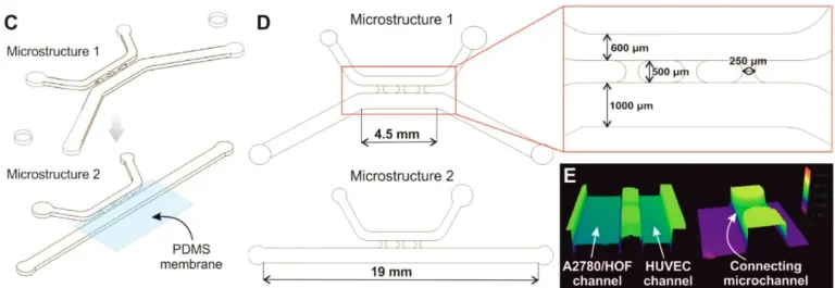
Figure 1. Channel layout and target dimensions. The two molded microstructures and their key measurements (1000 µm and 600 µm channels, 500 µm connectors), with a 3D laser scan (panel E) confirming the printed mold profile transferred into the PDMS cast. Source: Bazaz et al. Rapid Softlithography Using 3D-Printed Molds. Adv Mater Technol. 2019;4(10).
The thin membrane between the cancer and stromal channels was made separately, spin-coated onto a dish, cured, and cut to size, then bonded between the two molded layers using oxygen plasma activation. The result is a sandwich: fibroblasts seeded on one face of the membrane for the stromal channel, cancer cells on the other for the cancer channel, so the two populations sit in genuine layered contact, just as epithelial and stromal tissue do in the body.
Building the blood vessel used a clever trick called viscous finger patterning. The vascular channel was filled with liquid collagen, then a droplet of buffer was pushed through the still-soft gel. The flowing buffer carved a smooth cylindrical tunnel straight down the middle of the collagen, leaving a hollow lumen lined later with endothelial cells (HUVECs). Because the channel geometry was fixed by the printed mold, this self-formed vessel came out at a consistent, vessel-like diameter.
Cell loading was done in two timed steps with the chip flipped partway through, so each cell type settled onto the correct surface of the membrane and lumen. Over a 10-day culture, the team tracked cell health, vessel shape, cell migration through the connecting microchannels, VEGF secretion, and several genes tied to vessel growth and inflammation. A 3D laser scan of the molded cast confirmed the printed channel profiles transferred faithfully into the PDMS.
Results
The build worked, and it stayed alive. Across the full 10-day culture, every cell type held high viability (roughly 98–100%), confirming the molded device and its bonding chemistry were not harming the cells. The molded geometry transferred cleanly: the two stacked culture channels stayed separated by the 50–56 µm membrane, giving the layered tumor-and-fibroblast architecture the design was built around.
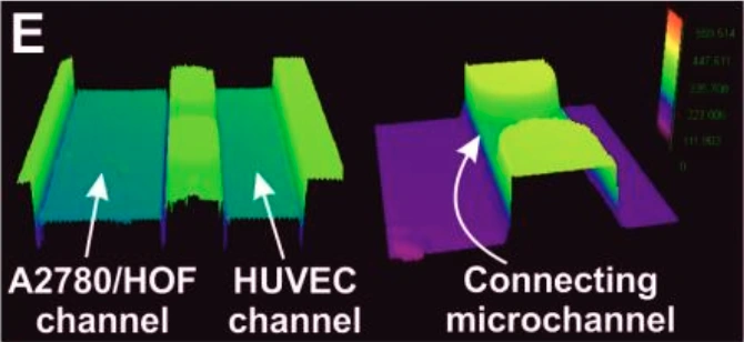
Figure 2. 3D laser scan of the cast surface, showing the molded channel walls for the tumor, vessel, and connecting microchannels reproduced from the printed mold. Source: Flont et al. Sensors. 2026;26:1503.
The self-formed vessel matched its target. The collagen lumen produced an average diameter of 370 ± 9 µm, putting it in the size range of small veins in the body, and a permeability check confirmed the vessel wall held together rather than leaking. The layered tumor construct itself measured 189 ± 17 µm thick, a genuine multilayer rather than a flat sheet.
The model also behaved like living tumor tissue. Endothelial cells did not sit still: over 60 hours they migrated more than 200 µm out of the vessel and toward the tumor layer, the hallmark of early angiogenesis. That movement was driven by signaling, the most complex co-culture pumped out the most VEGF, climbing past 5000 pg/mL by day 10, more than 4800 pg/mL above the day-1 baseline. Gene readouts backed this up, with vessel-growth genes (ANG, ANGPT2) and an inflammation gene (IL-6) rising over the culture, and fibroblasts shifting toward the activated, tumor-supporting phenotype seen in real disease. Together these results show a molded, castable device can reproduce not just the structure of a vascularized tumor but its dynamic behavior.
“This unique multicellular integration within a single system enabled, for the first time, the laboratory reconstruction of a 3D, heterogeneous, and vascularized ovarian cancer model.”
— Flont et al., Sensors, 2026
Products Used In This Study

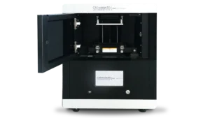
Questions on how our 3D printing system can work for your research? Connect with one of our specialists.
More Research Article Summaries
McGill University researchers build a 3D printed double-sided mold system to shape and merge brain organoids on a chip
University of Hawaiʻi researchers 3D print wearable "sweatainer" devices with true microscale internal channels
Want to read more article summaries?
We summarize the latest microfluidics and 3D printing research.
Curated from peer-reviewed publications featuring the CADworks3D system.