
Research Article Summary
5 Minute Read
McGill University researchers build a 3D printed double-sided mold system to shape and merge brain organoids on a chip
Title
Microfabricated dynamic brain organoid cocultures to assess the effects of surface geometry on assembloid formation
Authors
Camille Cassel de Camps, Sabra Rostami, Vanessa Xu, et al.
Journal
Biotechnology Journal, 2024; 19:e2400070
DOI Link
Title
Microfabricated dynamic brain organoid cocultures to assess the effects of surface geometry on assembloid formation
Authors
Camille Cassel de Camps, Sabra Rostami, Vanessa Xu, et al.
Journal
Biotechnology Journal, 2024; 19:e2400070
DOI Link
Summary
McGill researchers developed a 3D printed double-sided molding technique that casts two-piece PDMS microfluidic chips, letting them culture two brain organoid types separately, shape their surfaces, and then merge them into an assembloid on demand, with a design-to-device turnaround of under 8 hours. Cassel de Camps et al.
Summary Author
This page was prepared by CADworks3D to summarize and highlight a peer-reviewed research article authored by independent researchers utilizing the CADworks3D system.
Key Results at a Glance
<8 hours
Design-to-device turnaround
From CAD file to a usable cast PDMS chip in under one workday
~200 µm
Thinnest printed barrier
Double-sided molds produced separating walls as thin as ~200 µm
7 days
Confirmed cell viability
Stem cells and midbrain organoids both stayed viable at least a week in-device
2-piece
Interlocking mold design
Base + lid molds enabled double-sided features impossible with single-side casting
Objective
Organoids are three-dimensional tissue cultures grown from stem cells. Under the right conditions, these cultures differentiate toward diverse cell lineages and capture some of the complexity of real tissues. Multiple organoid types can also be assembled so they interact, fuse, and mature, and these combined structures are called assembloids. Because they contain more than one organoid type, assembloids capture additional cellular diversity and architectural complexity compared to single-type organoids. In the developing brain, complex circuits are established by neurons projecting and migrating to create local and long-distance connections, so regionalized brain organoids can be assembled to model the circuits that run throughout the brain.
The paper notes that the process by which organoids integrate to form an assembloid remains unclear, and may play an important role in the resulting structure. Tissue geometry is well established to influence fundamental cellular processes such as proliferation, differentiation, branching, and invasion. While previous studies have shown that geometric confinement drives the organization of developing neural structures, whether these geometric features play a role in assembloid formation was an open question.
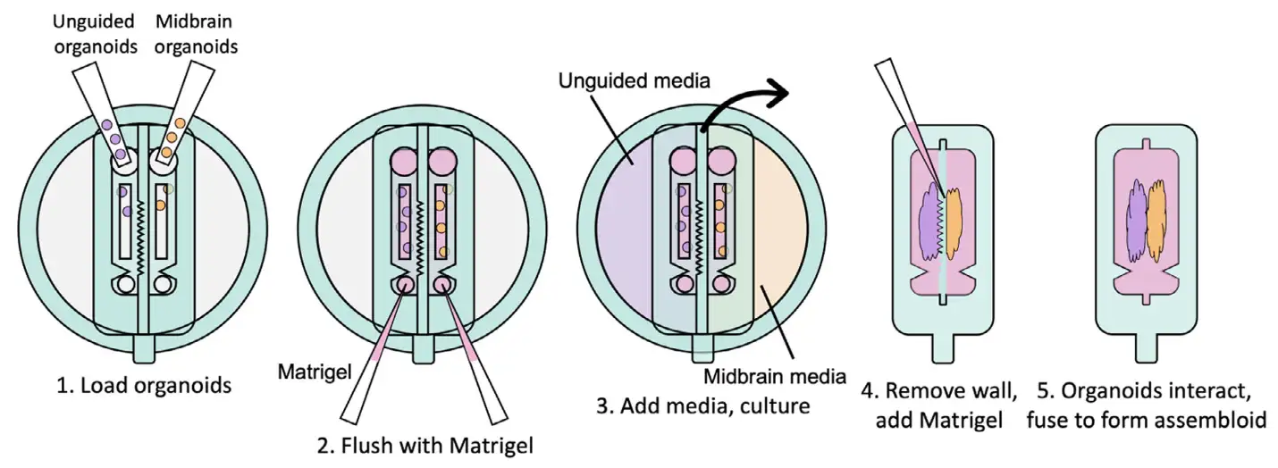
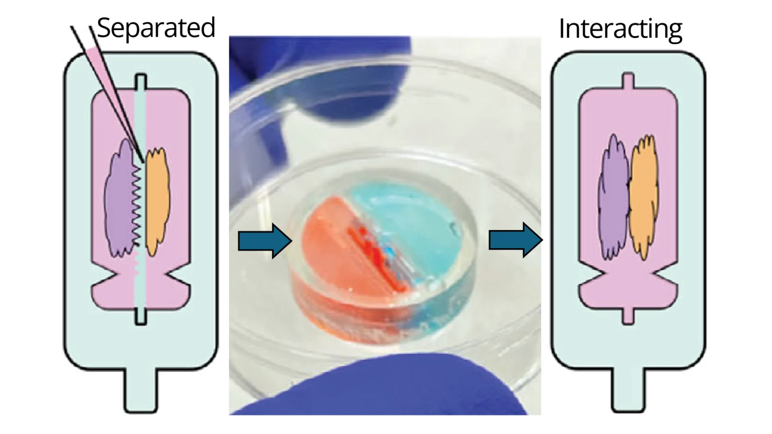
Figure 1.The device concept, shown step by step. Two different organoid types are loaded into separate channels, each fed by its own media reservoir. The shaped wall between the channels molds the organoid surfaces as they grow, and is then physically removed so the two organoids can meet, interact, and fuse into a single assembloid. Source: Cassel de Camps et al. Microfabricated dynamic brain organoid cocultures to assess the effects of surface geometry on assembloid formation. Biotechnology Journal. 2024.
Answering it requires specific technical capacity: a way to impose a chosen geometry on independently cultured organoids, control their relative positions before they interact, and support long-term culture in a format that keeps the tissue alive and visible. The work builds on a recent approach by Park et al., who used silicone inserts to restrict the shape of intestinal organoids as they grew, controlling geometry while supporting maturation. Inspired by it, the authors set out to culture distinct brain organoid types in adjacent compartments while shaping their surface geometry, then remove a separating insert so the positioned organoids could begin forming an assembloid. Supporting this required complex three-dimensional geometries with overhanging, double-sided features that could only be achieved through interlocking surfaces for double-sided PDMS molding, rather than conventional single-side replica molding.
Methodology and Design
The fabrication process used in the study begins with a mold that is designed and 3D printed, then washed and UV-cured to prepare it for casting. The device itself is cast in polydimethylsiloxane (PDMS), which is poured over the mold and degassed to remove trapped air before curing. Once cured, the PDMS is de-molded to release the finished piece from the printed mold. The surfaces are then treated to control how the organoids and matrix interact with them, sterilized, and assembled into a chip that supports coculture.
To evaluate how the platform supports organoid coculture and shapes assembloid formation, four core design features were tested:
Two-Channel Coculture Chip
Removable Insert Wall
Double-Sided Master Mold
Open-Slit Phase Guide
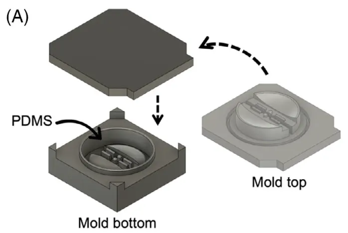
Figure 2. The two-piece displacement mold used to cast a device. The mold bottom and mold top each carry patterned features, and PDMS is shaped between them. Printing the mold in two halves is what lets a single cast part carry detail on both its top and bottom surfaces, which single-sided casting cannot do. Source: Cassel de Camps et al. Microfabricated dynamic brain organoid cocultures to assess the effects of surface geometry on assembloid formation. Biotechnology Journal. 2024.
The molds were designed in Fusion 360 and 3D printed at a layer thickness of 50 µm, then washed with isopropanol and cured in a 36 W UV chamber overnight. They were designed for assembly into chambers with patterned features on both the base and the lid, which is what enabled double-sided molding. The device itself was cast in polydimethylsiloxane (PDMS). PDMS prepolymer and curing agent were mixed at a 10:1 ratio by weight, poured into the chamber, and degassed under vacuum. The molded lid was lowered slowly from one side to avoid trapping air, then pressed down to displace excess PDMS, with tongue-and-groove features in the base and lid containing the prepolymer. The PDMS was cured overnight at 40°C to minimize shrinkage, then de-molded using 70% ethanol to release the devices from the 3D printed resin. Before culture, base devices were coated with dopamine to improve adhesion, while the removable inserts were passivated with Pluronic F-68 to reduce adhesion and allow clean release. Components were sterilized under UV and assembled on a coverslip that formed the bottom of the reservoirs.
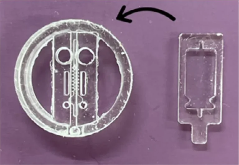
Figure 3. The replica-molded PDMS device after casting, with the base piece and the separate insert piece shown side by side. This is the physical chip produced from the printed molds, before the two pieces are assembled for culture. Source: Cassel de Camps et al. Microfabricated dynamic brain organoid cocultures to assess the effects of surface geometry on assembloid formation. Biotechnology Journal. 2024.
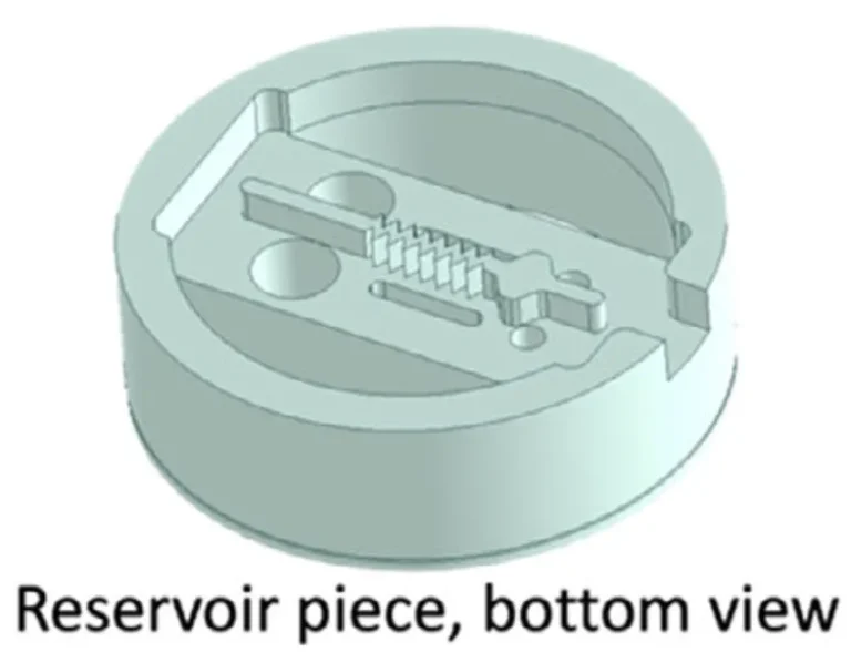
Figure 4. A 3D model of the removable insert, shown here with a triangular separating wall. This is the piece that shapes the organoid surface as it grows, and swapping in inserts with different wall geometries (flat, pointed, or mixed) is how the surface shape is chosen. The insert is later pulled out to let the organoids fuse. Source: Cassel de Camps et al. Microfabricated dynamic brain organoid cocultures to assess the effects of surface geometry on assembloid formation. Biotechnology Journal. 2024.
The cast devices were designed to allow pipetting of matrix and cells into the channels through inlet ports while leaving the channel tops open for nutrient exchange. This used an overhanging phase guide that holds injected liquid in place by surface tension, leaving a 600 µm slit open along the top for media exchange. Two adjacent channels were each fed by an independent media reservoir, so organoids with separate media requirements could be supported side by side. The dynamic co-culture devices were built as two pieces: a base holding the organoids, and an upper piece with the separating wall and reservoirs. The wall could be designed with different geometries; the authors tested flat versus triangular shapes. Pre-formed organoids were pipetted into loading ports, surrounded with matrix, and fed their appropriate media. Once the organoids had adopted the shapes defined by the compartment, the inserts were removed with tweezers, the gap was back-filled with matrix, and a combined differentiation medium was added, allowing the organoids to interact and fuse.
Results
The double-sided 3D printed molding chambers were essential, because the required overhanging, double-sided features could only be achieved through interlocking surfaces for double-sided PDMS molding. The authors state this method is extremely rapid and versatile, allowing design-to-device turnaround times of less than 8 hours, while creating structures that would be extremely difficult to produce with conventional single-side replica molding. The technique created barriers as thin as ~200 µm.
The team first verified the devices worked. Loading a model cell line stained two colours into adjacent channels (each 1 mm wide, separated by ~500 µm) and culturing for 3 days produced no colour exchange between compartments. When one reservoir was filled with dyed media, only cells in that channel were dyed, demonstrating functional reservoir separation.
The devices then supported living tissue at two stages. Stem cells loaded into the channels were confirmed viable for at least one week, and mature midbrain organoids likewise maintained viability for 7 days in culture, verified by live/dead staining. This confirmed the device could sustain cultures from the stem cell stage through to mature organoids. For assembloid formation, midbrain and unguided organoids were loaded into adjacent channels. After 5–28 days, the inserts were removed, leaving the organoids separated by the wall width (600 µm, variable by design). The organoids retained the wall’s shape after removal, immunostaining confirmed each organoid type expressed its expected distinct markers, and within 3 days the organoids bridged the gap to initiate assembloid formation.
Finally, the authors tested flat versus triangular separating-wall shapes. The organoids adopted the shapes provided by the wall, and after removal, cells migrated out regardless of geometry. However, migration distance differed by originating tissue shape: cells migrating from a flat organoid periphery travelled significantly farther than cells migrating from the flat midpoint of a triangular edge.
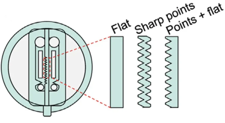
Figure 5. Schematic of the interchangeable separating walls, printed with different edge geometries, including flat, sharp points, and points combined with flat regions. Because the organoids grow against these walls, the chosen geometry sets the shape of the organoid surface being tested. Source: Cassel de Camps et al. Microfabricated dynamic brain organoid cocultures to assess the effects of surface geometry on assembloid formation. Biotechnology Journal. 2024.
“This fabrication method has the advantage of being extremely rapid and versatile, allowing design-to-device turnaround times of less than 8 hours, while creating novel structures that would be extremely difficult to produce using conventional single-side replica moulding approaches.“
— Cassel de Camps et al., Biotechnology Journal (2024)
Products Used In This Study

Questions on how our 3D printing system can work for your research? Connect with one of our specialists.
More Research Article Summaries
Sungkyunkwan University researchers build microfluidic blood-test discs from 3D printed molds at room temperature
University of Hawaiʻi researchers 3D print wearable "sweatainer" devices with true microscale internal channels
Want to read more article summaries?
We summarize the latest microfluidics and 3D printing research.
Curated from peer-reviewed publications featuring the CADworks3D system.