
Research Article Summary
5 Minute Read
UTS researchers build a lung-on-a-chip using 3D printed molds instead of a cleanroom
Title
A rapidly prototyped lung-on-a-chip model using 3D-printed molds
Authors
Jesus Shrestha, Maliheh Ghadiri, Melane Shanmugavel, et al.
Journal
Organs-on-a-Chip (Elsevier), Vol. 1, 2019, Article 100001
DOI Link
Summary
Shrestha et al. developed a rapid surface treatment protocol for 3D printed resin molds that lets researchers cast PDMS microfluidic chips without a cleanroom or specialist training. The team used the method to build and validate a functioning lung-on-a-chip device in under a day.
Summary Author
This page was prepared by CADworks3D to summarize and highlight a peer-reviewed research article authored by independent researchers utilizing the CADworks3D system.
Title
A rapidly prototyped lung-on-a-chip model using 3D-printed molds
Authors
Jesus Shrestha, Maliheh Ghadiri, Melane Shanmugavel, et al.
Journal
Organs-on-a-Chip (Elsevier), Vol. 1, 2019, Article 100001
DOI Link
Key Results at a Glance
24 hours
Mold-to-Chip Fabrication
CAD file to a bonded, ready-to-test chip, no cleanroom required
30 µm
XY Printing Resolution
Fine enough to reproduce the chip’s microchannel features accurately
Reusable molds
Repeated PDMS Casting
Surface-treated resin molds withstood multiple casting cycles without cracking
2 Week
Sustained Cell Viability
Lung epithelial cells stayed healthy and confluent in the chip for up to two weeks
Objective
Chronic respiratory disease is a major global health burden, and developing new drugs for it depends on lab models that behave like real lung tissue. The two most common laboratory setups, flat 2D cell cultures and transwell inserts, fall short here. Neither one reproduces the three-dimensional structure of airway tissue, the way neighboring cells communicate, or the air-exposed surface that lung cells actually experience in the body.
Organ-on-a-chip systems were developed to close that gap. These are small, PDMS (polydimethylsiloxane) based microfluidic devices that recreate the physical and functional conditions of an organ well enough to support drug screening and disease modeling. The problem is how they get made. The standard fabrication route, soft lithography, requires a cleanroom, specialized equipment, and a trained microfabrication technician. Turning around a single new mold design can take days, which makes it a poor fit for a biology lab that wants to test and adjust chip geometry quickly.
3D printing looked like an obvious shortcut for producing these molds faster and without a cleanroom. In practice, it has not been that simple. Most 3D printing resins leave a layer of uncured monomers and oligomers on the surface of a printed part, and that residue interferes with PDMS curing, so PDMS either will not release from the mold or will not fully cure against it. Existing workarounds for this exist, but they tend to be slow, inconsistent between labs, and not built for a mold that needs to be reused many times. This paper set out to fix that: build a faster, more reliable surface treatment protocol for 3D printed resin molds, then prove it works by using it to fabricate and validate a functioning lung-on-a-chip device.
Methodology and Design
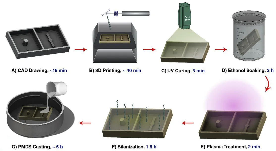
Fabrication starts with a CAD model of the mold, exported and printed on a resin-based digital light processing (DLP) 3D printer. The printed mold then goes through a short surface treatment (washing, UV post-curing, oxygen plasma treatment, and silanization) so that PDMS releases cleanly instead of sticking to the mold or failing to fully cure against it. The treated mold is used to cast two thin PDMS layers, which are trimmed, punched for inlets and outlets, and bonded around a porous membrane to form the finished chip.
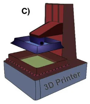
Figure 1. A digital light processing (DLP) 3D printer produces the resin mold used to cast the chip's PDMS layers. Source: Shrestha et al. A rapidly prototyped lung-on-a-chip model using 3D-printed molds. Organs-on-a-Chip. 2019.
That single (Open-Well Lung) chip design was then tested across four applications that reflect how a respiratory researcher would actually put it to use:
Barrier Model
Mucus Assay
Drug Transport
Smoke Exposure
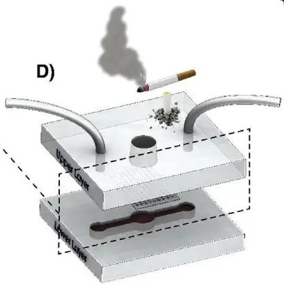
Figure 2. The finished chip design: an open-top well for cell seeding sits above a lower media channel, separated by a porous membrane. Source: Shrestha et al. A rapidly prototyped lung-on-a-chip model using 3D-printed molds. Organs-on-a-Chip. 2019.
On the printing side, feature size set the slice thickness: finer mold features were printed at 10 µm slices with a 1 second cure per layer, while coarser features used 30 to 50 µm slices, and a 24 second base layer anchored the print to the build platform. After printing, the mold was rinsed in isopropanol, post-cured under UV light for 3 minutes, then soaked in ethanol for 2 hours to strip away any remaining uncured monomer. A brief oxygen plasma treatment prepped the surface for silanization, which laid down a thin fluorinated coating so cured PDMS would peel away cleanly. PDMS was mixed at a 10:1 base to curing agent ratio, degassed, poured onto the treated mold, and cured at 45°C for 4 to 5 hours. Curing temperature mattered: higher temperatures cracked the mold surface and led to leaks later on. The two PDMS layers, with a porous polycarbonate membrane sandwiched between them, were plasma treated, aligned, bonded, then baked again and leak tested with food dye before use.
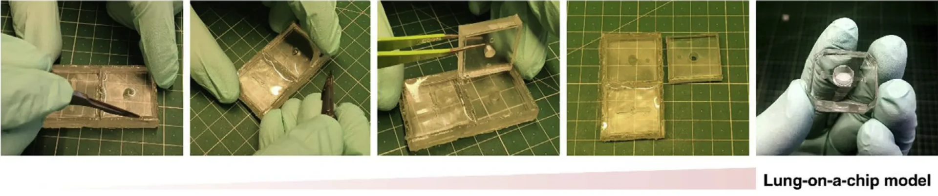
Figure 3. The 3D printed mold ready for PDMS casting (left) and the cured PDMS layer being peeled and prepared for bonding (right). Source: Shrestha et al. A rapidly prototyped lung-on-a-chip model using 3D-printed molds. Organs-on-a-Chip. 2019.
With the chip assembled, Calu-3 airway epithelial cells were seeded onto the membrane and grown at an air-liquid interface, meaning the top of the cell layer sat exposed to air rather than submerged in media, mimicking how airway tissue behaves in the body. For the barrier model, the team tracked how well the cell layer blocked a small fluorescent tracer, sodium fluorescein, from passing through, confirming the cells had formed tight, functional junctions. The mucus assay used Alcian blue staining to visualize mucus building up on the cell surface over several days, a sign of proper epithelial differentiation. For drug transport, the researchers measured surface expression of P-glycoprotein, a transporter protein that moves drugs across the epithelium, using flow cytometry. The smoke exposure application dosed the chip with cigarette smoke extract, with and without the anti-inflammatory drug Budesonide, to see whether the model could detect inflammatory and barrier changes relevant to respiratory disease research.
Results
The surface treatment held up in practice. Once treated, the 3D-printed molds released cured PDMS cleanly and supported repeated casting cycles without cracking or losing channel definition, something untreated resin molds could not manage. Paired with the printer’s 30 µm XY resolution, the process reproduced the chip’s microchannel features consistently from one mold to the next.
Turnaround was the biggest practical win over cleanroom soft lithography. From CAD file to a bonded, ready-to-test chip (printing, surface treatment, PDMS casting, and bonding included), the entire process fit inside a single working day. That timeline matters most for labs without cleanroom access or dedicated microfabrication staff who still want to iterate on chip design.
The chip performed biologically as well. Seeded with Calu-3 airway cells, it maintained a confluent, viable cell layer for up to two weeks, with cells forming tight junctions, secreting mucus, and expressing the drug transporter P-glycoprotein, all markers of a properly differentiated airway epithelium. When challenged with cigarette smoke extract, the model registered inflammatory and barrier changes consistent with published smoke-exposure studies, and those changes were dampened when cells were co-treated with Budesonide, indicating the chip is sensitive enough for toxicology and drug-response research.
“The ability to rapidly prototype these molds with little technical skills makes organ-on-a-chip modelling accessible to a broad group of researchers.”
— Shrestha et al. A rapidly prototyped lung-on-a-chip model using 3D-printed molds. Organs-on-a-Chip. 2019.
Products Used In This Study
Questions on how our 3D printing system can work for your research? Connect with one of our specialists.
More Research Article Summaries
McGill University researchers build a 3D printed double-sided mold system to shape and merge brain organoids on a chip
University of Hawaiʻi researchers 3D print wearable "sweatainer" devices with true microscale internal channels
Want to read more article summaries?
We summarize the latest microfluidics and 3D printing research.
Curated from peer-reviewed publications featuring the CADworks3D system.