
Research Article Summary
5 Minute Read
University of Virginia researchers 3D print a PDMS-free chip to track brain cancer cell migration
Title
Open-Top Patterned Hydrogel-Laden 3D Glioma Cell Cultures for Creation of Dynamic Chemotactic Gradients to Direct Cell Migration
Authors
Aditya Rane, Steven Tate, Jenna L. Sumey, et al.
Journal
ACS Biomaterials Science & Engineering, 2024, Vol. 10, pp. 3470–3477
DOI Link
Summary
Researchers at the University of Virginia built an open-top, PDMS-free microfluidic device, patterned using a 3D printed mold, to create sustained chemical gradients that guide glioma cell migration inside a living 3D hydrogel culture. (Rane et al.)
Summary Author
This page was prepared by CADworks3D to summarize and highlight a peer-reviewed research article authored by independent researchers utilizing the CADworks3D system.
Title
Open-Top Patterned Hydrogel-Laden 3D Glioma Cell Cultures for Creation of Dynamic Chemotactic Gradients to Direct Cell Migration
Authors
Aditya Rane, Steven Tate, Jenna L. Sumey, et al.
Journal
ACS Biomaterials Science & Engineering, 2024, Vol. 10, pp. 3470–3477
DOI Link
Key Results at a Glance
15 mm
Continuous Hydrogel-to-Channel Interface
Longest uninterrupted, post-free boundary achieved between the open hydrogel and its fluid channel
1 mm
Full-Depth Gradient Uniformity
Chemical gradients stayed consistent through the hydrogel’s entire depth, not just at the surface
48 hours
Sustained, Drift-Free Culture
Gradient held steady for two full days without the hydrogel drying out
7.5 µL/min
Continuous Flow Rate
Steady pump-driven flow tuned to match natural interstitial flow rates in brain tissue
Objective
Glioblastoma is the most common and aggressive form of primary brain cancer in adults, and one of the reasons it stays so hard to treat is how easily it spreads through healthy brain tissue. Understanding what drives glioma cells to migrate away from a tumor site could help researchers design better lab models and, eventually, therapies that keep cancer cells contained or steer them toward drugs meant to eradicate them.
Most lab models used to study this kind of migration rely on microfluidic chips: small networks of channels that can deliver a steady chemical gradient to guide cell movement in a dish. These chips are usually built from poly(dimethylsiloxane), or PDMS, a flexible silicone material that molds easily into fine channels. The problem is that PDMS leaches small molecules into the culture and absorbs others out of it, which distorts the very chemical gradients researchers are trying to study. PDMS channels are also typically shallow, often under 0.1 mm deep, so cells spend more time interacting with hard channel walls than with the soft, three-dimensional hydrogel meant to mimic the brain’s tumor microenvironment.
The research team set out to close this gap with a hydrogel culture platform that removes PDMS from the final device altogether, while staying deep enough (about 1 mm) for cells to experience a genuine 3D environment, and precise enough to deliver a controllable, sustained chemical gradient across the width of the culture.
Methodology and Design
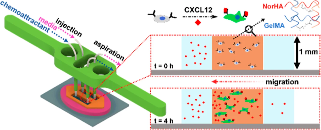
To build the device, the team first cast a silicone mold from a 3D-printed master mold, then used that silicone mold to shape a hydrogel directly on a glass slide. Once the hydrogel cured under UV light, the silicone mold was peeled away, leaving a patterned block of hydrogel sitting inside open channels rather than sealed tubing. A separate 3D-printed holder was then fitted onto the setup to connect the open channels to fluid lines, one delivering fresh media and chemical cues, the other pulling waste away, so the hydrogel’s exposed surface, not a plastic wall, became the interface where chemical gradients formed.
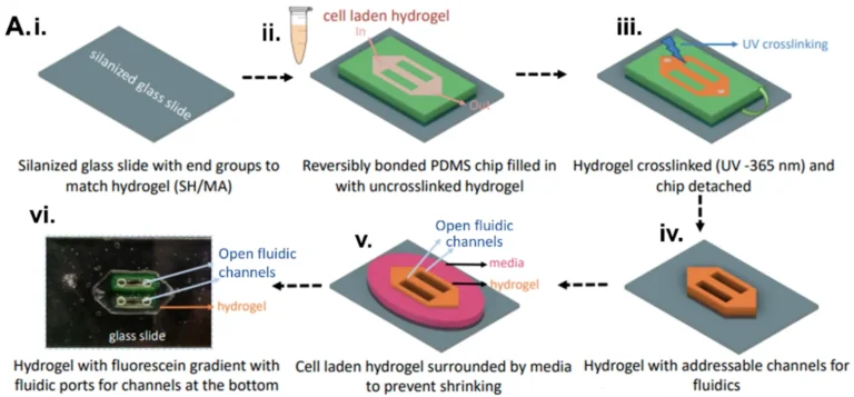
Figure 1. The hydrogel is cast and UV-cured inside a silicone mold on a glass slide. Once the mold lifts away, the patterned hydrogel is left with open channels bordering its surface, ready for fluid connections. Source: Rane, Tate, et al. Open-Top Patterned Hydrogel-Laden 3D Glioma Cell Cultures for Creation of Dynamic Chemotactic Gradients to Direct Cell Migration. ACS Biomaterials Science & Engineering. 2024.
This open, PDMS-free platform was then put to work in three ways to confirm it could support and study directed cell migration:
Small-Molecule Tracer Model
U87 Glioma Hydrogel Culture
CXCL12 / AMD3100 Assay
For gradient mapping, the team flowed two sizes of a fluorescent tracer through the open channels and tracked how each spread across the hydrogel over time: one sized to mimic a small-molecule drug (400 Da) and one closer to the size of a protein or cytokine (10 kDa). The small tracer reached a steady gradient within about an hour, while the larger one took up to 12 hours, showing that gradient speed through the platform naturally matches the diffusion behavior of different molecule classes.
For the migration assay, U87 glioma cells were embedded in a hybrid hydrogel made of hyaluronic acid to mimic the brain’s natural tumor environment, and gelatin methacrylate (GelMA), to give cells something to grip and migrate along. This hybrid formulation was chosen after comparing viability and migration across single-component hydrogels, and it kept cells healthy while still letting them align and move directionally across the patterned width.
For the drug response test, the chemokine CXCL12 was used to trigger calcium signaling and draw glioma cells toward the injection side of the channel, while a second set of cells was pretreated with AMD3100, a compound known to block that same signaling pathway. Cells exposed to CXCL12 alone showed a clear rise in calcium signal near the channel boundary, while AMD3100-pretreated cells showed almost no response, confirming the open platform can be used to test how well a candidate drug blocks a specific migration signal.
On the fabrication side, the hydrogel and channel pattern were kept to a length-to-width ratio near 2 mm and a length-to-height ratio near 1 mm, at or below a 5-to-1 aspect ratio overall, which let the silicone mold release cleanly from the cured hydrogel without tearing its edges. UV crosslinking ran at 5 mW/cm² for 120 seconds to fully cure the hydrogel while keeping cells viable, and glass slides were chemically treated beforehand so the finished hydrogel would bond to the glass instead of the mold. The 3D printed holder was produced in a clear, microfluidics grade resin so the culture could still be imaged with transmitted light, and it set the injection tubing below the hydrogel’s surface with the aspiration tubing level with it, using surface tension alone, no glue or clamps, to keep the open channel sealed. A pressure pump and vacuum line, run at a steady 7.5 µL/min, replaced the pulsing flow of a peristaltic pump, keeping the gradient stable across the full 48-hour culture period.
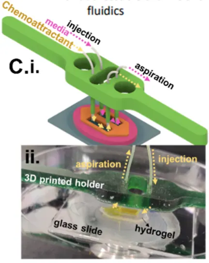
Figure 2. A 3D printed holder positions the injection and aspiration tubing against the open hydrogel channels, replacing the glued PDMS connections used in closed microfluidic chips. Source: Rane, Tate, et al. Open-Top Patterned Hydrogel-Laden 3D Glioma Cell Cultures for Creation of Dynamic Chemotactic Gradients to Direct Cell Migration. ACS Biomaterials Science & Engineering. 2024.
Results
The open-top design held up well across every test. The hydrogel-to-channel interface stayed continuous and uninterrupted across a full 15 mm length, with no support posts needed to keep the hydrogel in place, something closed systems typically require and which tends to create gaps in the interface.
Gradient uniformity was confirmed through the entire 1 mm depth of the hydrogel, not just at the surface, meaning cells anywhere within the 3D culture experienced a consistent signal rather than one dominated by contact with a channel wall. The gradient itself extended across a hydrogel width of about 2 mm and remained sustained for the full 48-hour culture window without the hydrogel drying out, a common failure point in open or PDMS-based systems.
Cell viability data supported the hybrid hydrogel formulation, with the HA-GelMA mix holding roughly 75% viability over 48 hours, notably higher than GelMA alone, while still letting glioma cells migrate at speeds comparable to the more permissive GelMA-only condition. In the drug response assay, CXCL12 stimulation produced a 1.8-fold increase in calcium signal over control, an increase that was effectively eliminated in cells pretreated with AMD3100, confirming the platform’s sensitivity to both migration cues and their inhibition.
“This hydrogel-based open fluidic system to deliver chemoattractant cues over square-centimeter-scale areas and millimeter-scale depths can potentially serve as a robust screening platform to assess emerging glioma models and chemotherapeutic agents to eradicate them.”
— Rane et al., ACS Biomaterials Science & Engineering, 2024
Products Used In This Study

Questions on how our 3D printing system can work for your research? Connect with one of our specialists.
More Research Article Summaries
McGill University researchers build a 3D printed double-sided mold system to shape and merge brain organoids on a chip
University of Hawaiʻi researchers 3D print wearable "sweatainer" devices with true microscale internal channels
Want to read more article summaries?
We summarize the latest microfluidics and 3D printing research.
Curated from peer-reviewed publications featuring the CADworks3D system.