
Research Article Summary
5 Minute Read
University of Virginia Researchers Build a Head-to-Head Guide for 3D Printing Microfluidic Devices
Title
Applied tutorial for the design and fabrication of biomicrofluidic devices by resin 3D printing
Authors
Hannah B. Musgrove, Megan A. Catterton, Rebecca R. Pompano
Journal
Analytica Chimica Acta, 2022
DOI Link
Summary
Musgrove et al. put three widely used 3D printing resins through head-to-head testing to show how print resolution, cell safety, and optical clarity are shaped by resin choice, printer settings, and part design.

Summary Author
This page was prepared by CADworks3D to summarize and highlight a peer-reviewed research article authored by independent researchers utilizing the CADworks3D system.
Title
Applied tutorial for the design and fabrication of biomicrofluidic devices by resin 3D printing
Authors
Hannah B. Musgrove, Megan A. Catterton, Rebecca R. Pompano
Journal
Analytica Chimica Acta, 2022
DOI Link
Key Results at a Glance
0.2 mm
Finest Channel Resolved
The smallest internal microchannel printed cleanly, using a low-viscosity resin under a 385 nm light source
~10x Lower Viscosity
Faster Channel Drainage
The high-resolution resin drained uncured material out of channels far more easily than standard clear resin.
> 60% Viability
Cell-Safe Contact at 4 Hours
Treated print material supported healthy primary immune cells after direct physical contact.
3 Resin Types
Head-to-Head Comparison
Biocompatible, optically clear, and high-resolution resins were benchmarked side by side.
Objective
Microfluidic devices, chips with channels smaller than a millimeter, let researchers run entire lab assays on a chip the size of a coin, using a fraction of the reagents, cells, and time required by benchtop methods. For over a decade, the standard way to build these devices has been soft lithography, casting a silicone polymer called PDMS over a hard mold. This works well for flat, two-dimensional designs, but complex parts require manually stacking, aligning, and bonding multiple PDMS layers by hand, a slow process highly sensitive to dust outside a cleanroom.
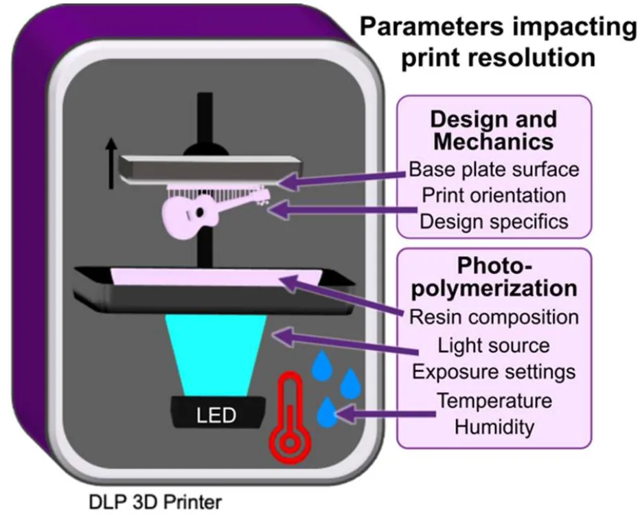
Resin 3D printing offers a way around these problems. Digital light processing, or DLP, printers cure a photopolymer resin one layer at a time using UV or violet light, building complex 3D structures as a single monolithic piece with no manual assembly. Interest in this approach has grown quickly, but labs adopting it run into familiar obstacles: commercial resins often cannot resolve the small channels a device needs, many contain components toxic to cells, and few are optically clear enough for microscopy. Guidance exists, but is scattered across dozens of papers and rarely compared side by side.
Musgrove et al. set out to close that gap, building a practical, data-supported reference that labs new to 3D printing could use to choose a resin, adjust print settings and part designs, and treat finished parts, to get reliable, cell-safe, optically clear devices without years of trial and error.
Methodology and Design
Every device in this study followed the same basic fabrication pipeline. Parts were designed as 3D models in CAD software, exported, and loaded into a slicing program that converts the design into printer-readable layers. A DLP printer then cured the resin layer by layer, building the part from the bottom up. Each finished piece was rinsed in isopropyl alcohol to remove uncured resin, dried, and exposed to additional UV light to fully harden, or post-cure.
To figure out which resin, setting, and design choices mattered most, the team built several purpose-made test devices, each one isolating a different part of the fabrication process.
Microchannel Test Chip
Reservoir Well Chip
Cell Culture Disk
Optical Test Disk
The microchannel test chip carried six internal channels with square cross sections from 0.2 to 1.2 mm, designed to reveal the smallest channel a given setup could keep open. The team printed copies using two light sources, 405 nm and 385 nm, and two resins with very different viscosities, then compared how cleanly the channels opened. They also varied layer height between 50 µm and 100 µm, and repositioned channels so fewer resin layers had to cure directly above them, a factor known as overhang.
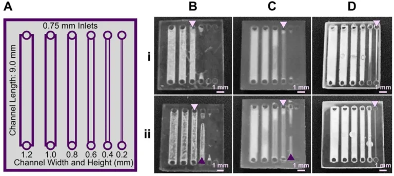
Figure 1. (A) The test piece design: six internal channels of decreasing size. (B) FormLabs Clear resin printed under a 405 nm light source. (C) The same resin under a 385 nm light source, resolving smaller channels than panel B. (D) A low-viscosity resin (BV007a), also printed at 385 nm, resolving the smallest channels of the three. Source: Musgrove et al. Applied tutorial for the design and fabrication of biomicrofluidic devices by resin 3D printing. Analytica Chimica Acta. 2022.
The reservoir well chip focused on a different failure mode: cracking. Hollow, cup-shaped features like wells and ports are prone to a defect called cupping, where a vacuum forms inside the hollow space as each cured layer peels away from the resin vat’s film, and the pressure difference cracks the part. To fix this, the team printed well designs with progressively thicker walls, filleted bases, and fully rounded exterior corners in place of the original thin-walled, sharp-cornered design, then checked each version for cracks and pinholes.
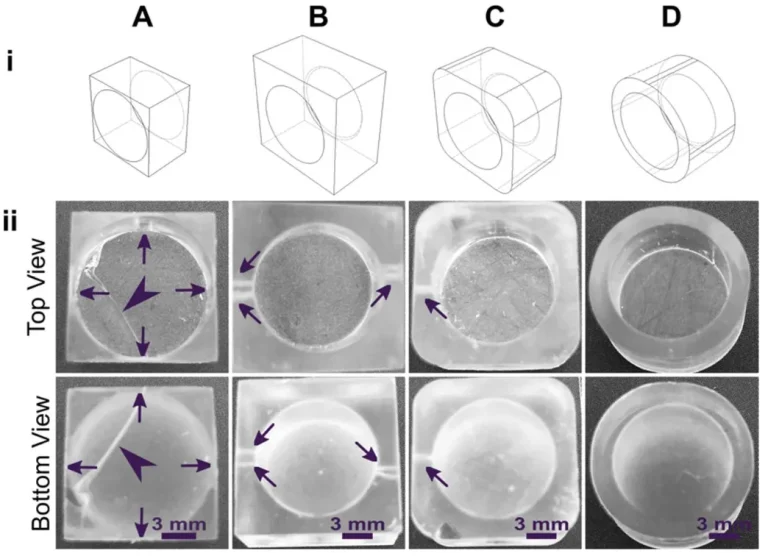
Figure 2. (A) The original well design: thin walls and sharp 90-degree corners, prone to cracking. (B) Thicker walls and a filleted base reduced cracking but still left small pinholes. (C) Partially rounding the outer corners reduced the strain further. (D) Fully rounding the outer corners eliminated cracks and pinholes entirely, the strongest design tested. Source: Musgrove et al. Applied tutorial for the design and fabrication of biomicrofluidic devices by resin 3D printing. Analytica Chimica Acta. 2022.
For the cell culture disk, 15 mm wide, 1 mm thick disks were printed in each test resin, then split into treated and untreated groups. Treated disks were soaked in sterile saline for 24 hours at 37°C or 50°C, depending on the resin, to leach out unbound, potentially toxic components before touching living cells. A second batch compared four post-treatment routes, saline soak, incubation, and autoclaving, to see which cleared toxins most effectively. All disks were rinsed, dried, UV sanitized, and cultured with primary mouse immune cells.
The optical test disk covered transparency and background glow. Thin disks were imaged under four microscopy filter sets against a PDMS control to measure autofluorescence, while thicker disks were tested with five post-processing methods: printing on glass, nail polish coating, resin coating, sanding, and buffing, each benchmarked against a glass slide.
Results
Resin choice and light source had the single biggest impact on channel resolution. Under a 405 nm light source, the standard clear resin only opened channels down to 0.6 mm, but switching to a 385 nm source, which better matched the resin’s light-absorbing chemistry, resolved channels roughly 0.2 mm smaller. The high-resolution resin performed even better. With a viscosity of only 75 to 100 mPa·s, about 10 times lower than the standard clear resin, it drained uncured material out of channels almost completely on its own, resolving channels as small as 0.2 mm, the finest size tested, at 385 nm and a 50 µm layer height.
Design changes mattered nearly as much as resin selection. Doubling layer height from 50 µm to 100 µm modestly improved resolution, and cutting overhang layers from 30 down to 10 improved it by about 0.2 mm on its own. For hollow features, replacing sharp 90-degree corners with fully rounded exterior walls eliminated cracking and pinholes by spreading shrinkage stress evenly.
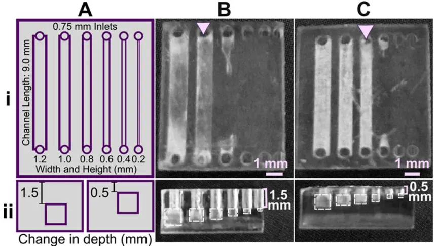
Figure 3. (A) Schematic of the test piece's channel layout, shown from the top and side. (B) Channels printed with a thick 1.5 mm overhang, 30 resin layers curing directly above the channel, resolving down to 1.0 mm. (C) The same design with the channel repositioned closer to the surface, a thin 0.5 mm overhang of just 10 layers, resolving channels about 0.2 mm smaller. Source: Musgrove et al. Applied tutorial for the design and fabrication of biomicrofluidic devices by resin 3D printing. Analytica Chimica Acta. 2022.
Biocompatibility depended heavily on resin type and post-treatment. After a 24-hour saline soak, primary immune cells stayed above 60% viability in direct contact with the treated biocompatible and standard clear resins for 4-hour experiments, not significantly different from the untreated plate control. The high-resolution resin, with its higher percentage of additives, remained largely toxic even after treatment. Heat and saline soaking were the most effective post-treatment methods overall, while autoclaving was faster but slightly less effective. Viability for all treated materials declined by 24 hours, pointing to leaching as a better fit for short experiments than extended culture.
For optical performance, the high-resolution resin showed negligible autofluorescence across all four channels tested, closely matching PDMS, while the two other resins glowed noticeably under UV excitation. Clarity came down to surface finish rather than resin chemistry. A simple coat of clear nail polish brought all three resins to a transparency statistically indistinguishable from a glass slide, the fastest practical fix for imaging-ready parts.
“In using 3D printing for production of microfluidic devices, compromises and strategic design choices are often required to best match the material and design to the required experiment.”
— Musgrove et al., University of Virginia, 2022
Products Used In This Study

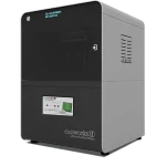
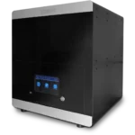
Questions on how our 3D printing system can work for your research? Connect with one of our specialists.
More Research Article Summaries
McGill University researchers build a 3D printed double-sided mold system to shape and merge brain organoids on a chip
University of Hawaiʻi researchers 3D print wearable "sweatainer" devices with true microscale internal channels
Want to read more article summaries?
We summarize the latest microfluidics and 3D printing research.
Curated from peer-reviewed publications featuring the CADworks3D system.