
Research Article Summary
5 Minute Read
University of Toronto researchers 3D print a microfluidic chip to catch rogue stem cells hiding in lab-grown heart tissue

Title
Ultrasensitive and rapid quantification of rare tumorigenic stem cells in hPSC-derived cardiomyocyte populations
Authors
Zongjie Wang, Mark Gagliardi, Reza M. Mohamadi, et al.
Journal
Science Advances, Vol. 6, Issue 12, eaay7629 (2020)
DOI Link
Summary
Wang et al. built a low-cost, 3D printed microfluidic chip that can find five rogue stem cells hiding among one million lab-grown heart cells, a detection limit current lab tools cannot match. The design relies on graded-height flow channels, made possible by a rapid 3D print-and-mold fabrication process instead of traditional cleanroom photolithography.

Summary Author
This page was prepared by CADworks3D to summarize and highlight a peer-reviewed research article authored by independent researchers utilizing the CADworks3D system.
Title
Ultrasensitive and rapid quantification of rare tumorigenic stem cells in hPSC-derived cardiomyocyte populations
Authors
Zongjie Wang, Mark Gagliardi, Reza M. Mohamadi, et al.
Journal
Science Advances, Vol. 6, Issue 12, eaay7629 (2020)
DOI Link
Key Results at a Glance
0.0005%
Detection Limit
Found 5 rogue stem cells hiding among 1 million lab-grown heart cells
30 x 30 µm
Print Resolution
Pixel size the 3D printer achieved to build the chip’s stepped flow channels
40 Chips/Day
Fabrication Output
Devices one researcher could mold from a single 3D printed master, per day
$4/Chip
Material Cost
Price per finished chip once the reusable 3D-printed mold was ready
Objective
Stem cell therapies derived from human pluripotent stem cells (hPSCs) are moving toward the clinic for conditions such as heart failure and neurodegeneration. To make a batch of heart muscle cells, called cardiomyocytes or CMs, researchers coax hPSCs to differentiate in culture. That process is never perfectly efficient. A small number of undifferentiated hPSCs can survive it and end up hiding inside the final CM population, unnoticed.
That leftover fraction matters because undifferentiated hPSCs keep the exact property that made them useful earlier in the pipeline: the ability to self-renew and turn into almost any cell type. Left inside a therapeutic cell product, that property becomes a liability. Animal studies have shown that hPSC contamination below 0.025 percent, a level under what older tools can reliably measure, is enough to trigger a teratoma, a tumor made up of multiple tissue types, after implantation. Before a batch of lab-grown heart cells can be considered safe for a patient, someone has to count how many rogue stem cells are hiding inside it, down to a fraction most instruments cannot resolve.
The two standard tools for this quality check were not built for the job. Flow cytometry loses accuracy at very low contamination levels because of sampling limits, and reliably detecting a population at 0.1 percent would require collecting an impractically large number of events per sample. PCR-based methods hit a different wall: the genetic signal from a handful of rare hPSCs gets buried under the much larger background signal from surrounding differentiated cells, along with artifacts introduced during reverse transcription.
That left a real gap in stem cell manufacturing quality control: a method sensitive enough to catch rare hPSCs at clinically meaningful concentrations, while staying fast and cheap enough to run on every batch. Wang et al. set out to close that gap with a microfluidic device that could be fabricated on demand rather than in a cleanroom, opening the door to a more practical, routine form of testing.
Methodology and Design
The core of the study is a single microfluidic chip, built through a rapid design and molding pipeline instead of traditional cleanroom photolithography. Researchers laid out the channel pattern digitally, 3D printed it as a positive master, cast a soft silicone material called polydimethylsiloxane, or PDMS, onto that master to form a reusable negative mold, then cast PDMS a second time to produce the finished chip layer, which was sealed onto a glass slide to close off the channels.
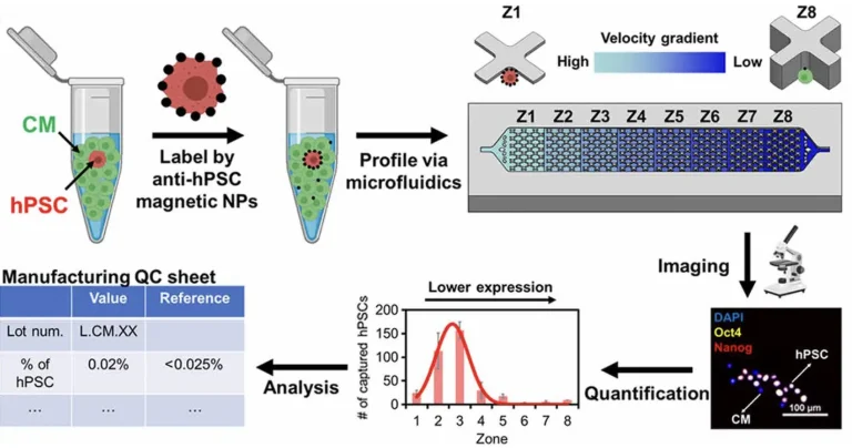
Figure 1. How the chip works, start to finish. Cells are magnetically labeled, sorted inside the 3D printed device, then imaged to generate a stem cell count for quality control. Source: Wang et al. Ultrasensitive and rapid quantification of rare tumorigenic stem cells in hPSC-derived cardiomyocyte populations. Science Advances. 2020.
That same chip design (SCQC Chip) was then put to work across three applications in the study, each testing a different part of what a manufacturing ready detection tool needs to do.
Rare Cell Detection
Tumor Risk Screening
Live Cell Recovery
For rare cell detection, cells were first labeled with magnetic nanoparticles that bind a surface marker found on hPSCs but not on CMs. This mixture was pumped into the chip, which contains eight sequential capture zones, each with a slightly taller channel than the last, creating a stepped flow velocity gradient from inlet to outlet. Cells carrying more magnetic label resist the drag of faster flow and get trapped in the earlier, high-velocity zones, while CMs with little or no label are swept into later zones or flushed out entirely. Imaging and counting the cells caught in each zone produces a profile showing how many hPSCs are present.
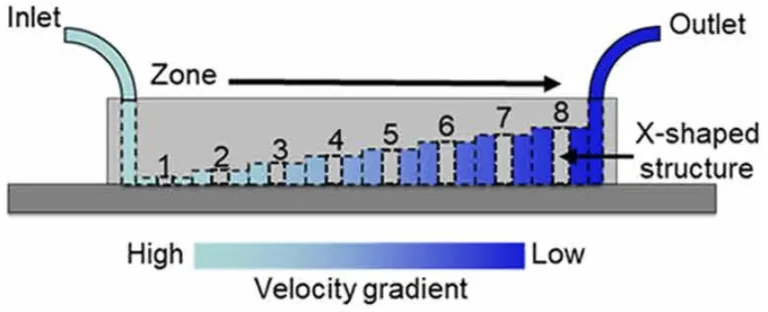
Figure 2. Inside the chip's design: eight stepped zones, each slightly taller than the last, create a flow velocity gradient that sorts cells by how strongly they are magnetically labeled. Source: Wang et al. Ultrasensitive and rapid quantification of rare tumorigenic stem cells in hPSC-derived cardiomyocyte populations. Science Advances. 2020.
For tumor risk screening, that same readout was generated on cardiomyocyte batches before they were injected into mice, and the chip’s contamination estimates lined up with which animals actually went on to grow teratomas, a comparison flow cytometry could not reliably make on the same samples. For live cell recovery, the magnetic field was switched off after capture so trapped hPSCs could be released intact, cultured back into colonies, and tested to confirm they still behaved like genuine stem cells.
A few fabrication details made this possible. The 3D printer used for the master mold was tuned to a pixel size of 30 by 30 µm, fine enough to resolve the eight stepped channel heights, each differing from its neighbor by 50 µm, that create the flow velocity gradient. A single printed master, produced in under an hour, could be reused to cast many PDMS negative molds, and each negative could cast multiple finished chips in turn, an approach that skips the mask alignment steps that normally slow down multi-height microfluidic fabrication. Sealing was handled with plasma bonding to glass coverslips followed by a brief oven bake, and each finished device was sandwiched between small magnet arrays with alternating polarity before being connected to a syringe pump to drive flow.
Results
The finished chip detected five rogue stem cells hiding in a background of one million differentiated heart cells, a limit of detection of 0.0005 percent. That is roughly two orders of magnitude more sensitive than flow cytometry and PCR-based methods, which topped out around 0.2 to 0.4 percent when tested side by side on the same samples. At the flow rate the team settled on, the chip captured 85.7 percent of spiked stem cells while flushing away 99.7 percent of the surrounding heart cells, the combination that makes such a low detection limit possible.
None of this would have worked without the fabrication side holding up its end. Printing the master mold at a 30 by 30 µm pixel size resolved the chip’s eight stepped capture zones, and because that single master could be reused to cast negative and positive PDMS molds repeatedly, one researcher could produce about 40 chips per day at a material cost of roughly $4 per chip, a steep drop from the cost and turnaround typically tied to cleanroom microfabrication.
The chip also proved gentle enough for downstream use. Rare hPSCs recovered live from the device grew back into colonies and kept the ability to differentiate into all three germ layers, confirming that sorting cells by flow velocity does not damage the cells the test is meant to protect.
“[The chip] provides an ultrasensitive, rapid, inexpensive, and scalable means of quantifying rare hPSCs.”
— Wang et al., Science Advances (2020)
Products Used In This Study

Questions on how our 3D printing system can work for your research? Connect with one of our specialists.
More Research Article Summaries
McGill University researchers build a 3D printed double-sided mold system to shape and merge brain organoids on a chip
University of Hawaiʻi researchers 3D print wearable "sweatainer" devices with true microscale internal channels
Want to read more article summaries?
We summarize the latest microfluidics and 3D printing research.
Curated from peer-reviewed publications featuring the CADworks3D system.