
Research Article Summary
5 Minute Read
University of Maryland researchers 3D print hollow microneedle arrays directly onto fluidic capillaries
Title
3D-Printed Microinjection Needle Arrays via a Hybrid DLP-Direct Laser Writing Strategy
Authors
Sunandita Sarker, Adira Colton, Ziteng Wen, et al.
Journal
Advanced Materials Technologies, 2023, Vol. 8, Article 2201641
DOI Link
Summary
Researchers built hollow, high-aspect-ratio microneedle arrays by combining two light-based 3D printing methods, printing the tiny needles directly onto 3D printed fluidic capillaries so the finished part needs no glue or adapters to connect to an injector. The approach produced needles with 30 µm inner diameters, 50 µm outer diameters, and 550 µm heights, opening a path to distributed microinjection into soft tissue (Sarker et al.).
Summary Author
This page was prepared by CADworks3D to summarize and highlight a peer-reviewed research article authored by independent researchers utilizing the CADworks3D system.
Title
3D-Printed Microinjection Needle Arrays via a Hybrid DLP-Direct Laser Writing Strategy
Authors
Sunandita Sarker, Adira Colton, Ziteng Wen, et al.
Journal
Advanced Materials Technologies, 2023, Vol. 8, Article 2201641
DOI Link
Key Results at a Glance
18
Capillaries per run
Two batches printed in a single DLP job
<45 min
Capillary print time
Full batch of fluidic bases printed in under 45 minutes
50µm Outer Diameter
Hollow needles
30 µm inner, 50 µm outer, 550 µm tall, spaced 100 µm apart
No adapters
Direct fluidic seal
Needles printed straight onto the capillary, no glue needed
Objective
Microinjection is one of the most common procedures in biomedical research, underpinning work from in vitro fertilization and drug delivery to developmental biology and genetically modified animal models. In nearly all of these settings, the standard tool is a single hollow needle that delivers its payload, whether cells, DNA, or nanoparticles, to one specific location. That works, but everything arrives at a single point.
For some applications, concentrating delivery at one spot creates problems. In stem cell therapy for the brain, a major obstacle is that donor cells often do not survive after injection. One reason is cell crowding: when a single needle deposits a high concentration of cells into one location, they clump together, and the cells trapped inside a clump lose easy access to oxygen and nutrients. Spreading the same payload across many smaller sites could ease that crowding, but that requires an array of needles rather than one.
Microneedle arrays are attractive because they distribute delivery over a wider area in a single step. The catch is that most existing arrays use solid needles, which cannot carry flowing liquid. For active fluid injection the needles must be hollow, and that is where manufacturing becomes the bottleneck. Building needles that are simultaneously hollow, thin, and tall (a high aspect ratio, meaning height much greater than width) is hard. Traditional cleanroom methods are slow and expensive and tend to produce only short, stubby shapes. Yet reaching the cerebral cortex of a mouse demands needles with outer diameters of tens of µm combined with heights above 500 µm. This paper sets out to close that gap, fabricating dense arrays of hollow, high-aspect-ratio needles and, just as importantly, connecting them to fluidic hardware by printing them directly onto their base, eliminating the manual gluing and alignment that limited earlier designs.
Methodology and Design
The work pairs two complementary light-based printing techniques. A digital light processing (DLP) printer builds the larger fluidic capillaries that carry liquid to the needles, while a separate direct laser writing step adds the microscopic needles directly on top of each capillary. The central idea is that the small needles are printed onto the larger printed base rather than made separately and attached later, so the two parts are fluidically sealed from the start.
To put this hybrid process into practice, three components were fabricated and evaluated, each serving a distinct role in the injection platform:
Hollow microneedle array
Fluidic capillary base
Microneedle-capillary assembly
The hollow microneedle array is produced through direct laser writing, a far higher-resolution technique than DLP printing. Rather than flashing whole layers, it focuses a pulsed infrared laser to a tiny point and scans it through the resin, curing material only where the laser is intense enough. That point-by-point control is what makes hollow needle features tens of µm wide possible, giving the array the fine geometry needed for fluid microinjection.
The fluidic capillary base is built during the DLP stage and shaped to plug directly into the target injector, removing the need for any separate adapter or sealant. This design lets the base serve directly as the injector interface once printed.
The microneedle-capillary assembly is formed by printing the needle array so that it begins slightly below the top surface of each capillary, overlapping into it rather than resting on top. This overlap fuses the needles to the base during printing, producing a single sealed part instead of two components joined afterward. The finished assembly is what is used to inject fluid into brain tissue.
The process begins in design software, where the team modeled batches of capillaries together. DLP printing uses a projector to flash patterns of UV light onto a tray of resin, curing one full layer at a time and building upward. Because an entire layer cures in a single flash, DLP is well suited to producing many capillaries at once, each held to its batch by five thin connecting struts. Printing every capillary in a fixed, predefined position also meant the whole batch could load into the direct laser writing system as a single unit, eliminating the by-eye alignment earlier methods required. After printing, the batch was developed in solvent, the capillary interiors were flushed to clear leftover resin, and the parts were rinsed and briefly UV-cured before moving to the laser writing stage.
After the needle arrays were written onto the capillaries, a final develop and dry step was performed, and individual assemblies were freed by cutting the five connecting struts, leaving a single sealed part ready for an injector.
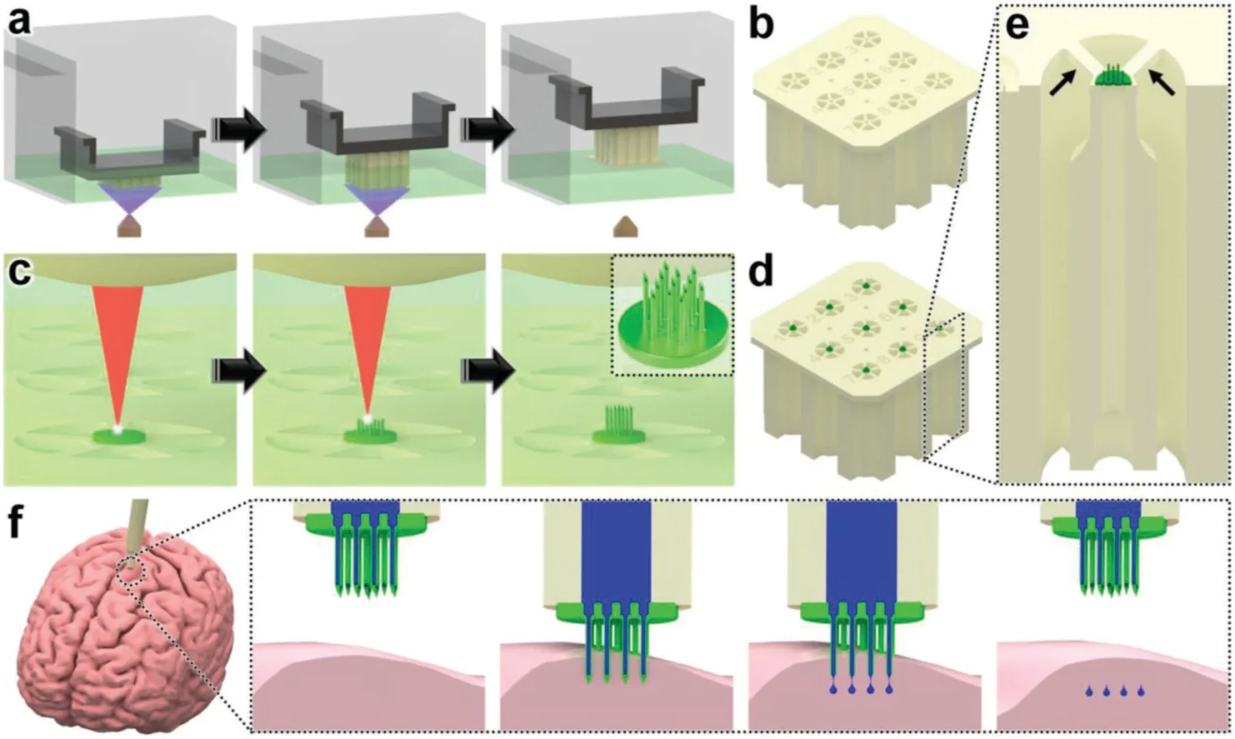
Figure 1. The two-stage hybrid printing workflow. First, DLP printing builds a batch of capillaries layer by layer (a, b). Next, direct laser writing prints hollow, high-aspect-ratio needle arrays straight onto each printed capillary, fusing them into one sealed part (c, d, e). Individual units are then released from the batch and used to inject fluid into brain tissue (f). Source: Sarker et al. 3D-Printed Microinjection Needle Arrays via a Hybrid DLP-Direct Laser Writing Strategy. Advanced Materials Technologies. 2023
Results
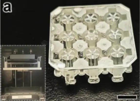
The fabrication process behaved as intended, and the parts came out clean. A single DLP run produced two batches totaling 18 capillaries in under 45 minutes, each cleanly attached by its five struts. DLP’s geometric control also gave each capillary a custom-shaped base that mated directly with the injector.
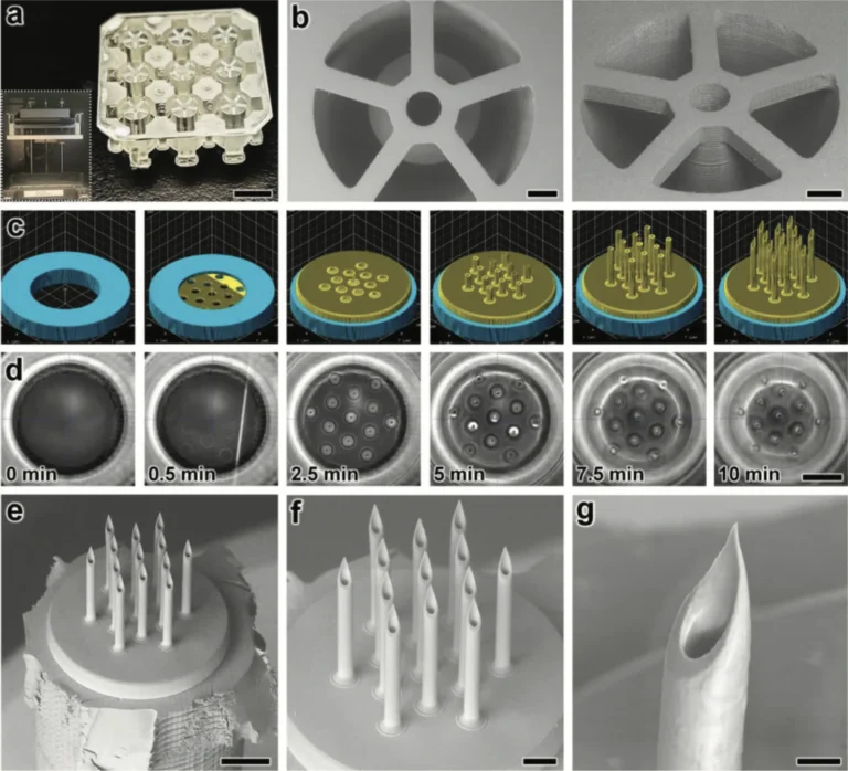
Figure 2. Fabrication results: a printed batch of capillaries (a), a close-up of one capillary and its struts (b), the CAD/CAM model and time-lapse of the needle print (c, d), and electron-microscope views of the finished hollow needles (e-g). Source: Sarker et al. 3D-Printed Microinjection Needle Arrays via a Hybrid DLP-Direct Laser Writing Strategy. Advanced Materials Technologies. 2023.
Under an electron microscope, the printed needles were well aligned and integrated with the capillary beneath them, with no visible defects along the needle-capillary interface. That interface matters most, because it is the joint where leaks or detachment would occur if the two-step strategy failed. The clean result is strong evidence that printing the needles directly onto the base, with that deliberate overlap, creates a genuine seal rather than a fragile contact.
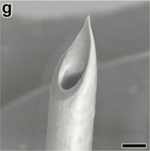
Figure 3. Close-up electron microscope view of a single printed needle tip. The open bore and thin, even wall show the submicron resolution the direct laser writing step can hold at the tip of a 550 µm tall needle. Source: Sarker et al. 3D-Printed Microinjection Needle Arrays via a Hybrid DLP-Direct Laser Writing Strategy. Advanced Materials Technologies. 2023
The laser writing step added the full array in about 10 minutes per capillary, producing the target geometry: 30 µm inner diameter, 50 µm outer diameter, 550 µm height, and 100 µm spacing. These numbers fall squarely in the range conventional manufacturing has struggled to reach, pairing a narrow width with a height many times larger. Close-up imaging showed the array survived manual release intact, tips and shapes preserved. Imaging after further handling told the same story, with no needles separating from the capillary. Together, the results show the hybrid strategy is a repeatable route to dense arrays of hollow, high-aspect-ratio needles that arrive already connected to fluidic hardware, with no glue, no adapters, and no manual alignment.
“The presented hybrid additive manufacturing strategy offers unique potential as an enabling technology for realizing entirely new classes of MNAs [microneedle arrays] to advance scientific discovery and promote human health and well-being”
— Sarker et al., Advanced Materials Technologies (2023)
Products Used In This Study
Questions on how our 3D printing system can work for your research? Connect with one of our specialists.
More Research Article Summaries
McGill University researchers build a 3D printed double-sided mold system to shape and merge brain organoids on a chip
University of Hawaiʻi researchers 3D print wearable "sweatainer" devices with true microscale internal channels
Want to read more article summaries?
We summarize the latest microfluidics and 3D printing research.
Curated from peer-reviewed publications featuring the CADworks3D system.