
Research Article Summary
5 Minute Read
University of Cambridge Researchers Build a 3D-Printed Model to Track Cochlear Implant Scarring in Real Time
Title
Tissue-Engineered Cochlear Fibrosis Model Links Complex Impedance to Fibrosis Formation for Cochlear Implant Patients
Authors
Simone R. de Rijk, Alexander J. Boys, Iwan V. Roberts, et al.
Journal
Advanced Healthcare Materials, Vol. 12, Issue 24, Article 2300732 (2023)
DOI Link
Summary
de Rijk et al. at the University of Cambridge engineered a 3D-printed bioreactor model that recreates the scar tissue forming around cochlear implants, and used it to uncover an electrical marker that can track that scarring directly from a patient’s own implant. The marker was then validated in a small group of recently implanted patients over five months.
Summary Author
This page was prepared by CADworks3D to summarize and highlight a peer-reviewed research article authored by independent researchers utilizing the CADworks3D system.
Title
Tissue-Engineered Cochlear Fibrosis Model Links Complex Impedance to Fibrosis Formation for Cochlear Implant Patients
Authors
Simone R. de Rijk, Alexander J. Boys, Iwan V. Roberts, et al.
Journal
Advanced Healthcare Materials, Vol. 12, Issue 24, Article 2300732 (2023)
DOI Link
Key Results at a Glance
60% ± 35%
Average Construct Contraction
The fibroblast-seeded gel, cast inside a 3D-printed mold around the implant electrode array, shrank substantially over 14 days as it built up scar-like tissue
Days 7-9
Electrical Inflection Point
Resistance in the model began climbing sharply in this window, marking the onset of measurable tissue buildup around the electrodes
30 µm
Mold and Platform Resolution
The casting mold and electrode-positioning platform were 3D printed at 30 µm XY resolution to keep the array precisely centered during tissue growth
4 Patients, 5 Months
Clinical Validation
The new electrical marker rose significantly in real cochlear implant patients over two postoperative check-ins, matching the lab model’s pattern
Objective
Cochlear implants restore hearing for people with severe sensorineural hearing loss by directly stimulating the auditory nerve with electrical pulses delivered through an electrode array placed inside the cochlea. They are one of the most effective neural prostheses in use today, but the body treats the implant as a foreign object. Mechanical trauma from insertion triggers an immune response: immune cells arrive first, followed by fibroblasts that lay down extracellular matrix (ECM) and gradually contract it into a fibrous capsule around the electrode array. In some patients, this capsule stays a thin sheath, and in others it progresses toward new bone formation, but either way it can dampen the electrical signal reaching the auditory nerve and contribute to residual hearing loss.
The trouble is that clinicians have no reliable way to track this scarring while it is happening. The only definitive method, histology, requires the tissue to be examined after death. Cochlear implant systems can report a contact “impedance,” but this is really a voltage reading at a single point in the stimulation pulse rather than a true electrical impedance measurement, and it varies significantly from person to person. Lab models exist too, but 2D cell cultures cannot recreate the way tissue deposits and contracts in three dimensions around an implant, and animal studies typically require the tissue to be examined after the animal is euthanized.
This study set out to close that gap by building a 3D, tissue-engineered model of cochlear fibrosis using a real, clinical-grade cochlear implant electrode array, then tracking its electrical behavior continuously as scar-like tissue formed and contracted around it. The goal was to identify an electrical signature of fibrosis that could eventually be read directly from a patient’s own implant, without additional surgery or equipment.
Methodology and Design
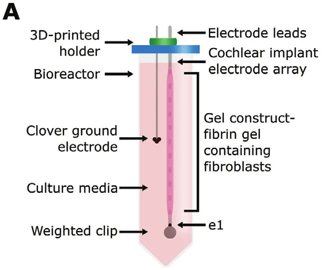
To recreate cochlear scarring in the lab, the team needed a way to grow fibrous tissue directly around a real implant electrode array while keeping everything in a fixed, repeatable position for electrical testing. They did this by casting a fibroblast-seeded fibrin gel around a clinical-grade electrode array inside a 3D-printed mold, then suspending the whole assembly inside a bioreactor for two weeks while the cells remodeled the gel into a denser, scar-like structure.
Three separate 3D-printed components made that process possible, each solving a specific positioning problem in the build:
Casting Mold
Electrode Platform
Wiring Holder
The casting mold held the fibrinogen and thrombin solution in place as it set around the electrode array, giving every construct the same starting shape and keeping the array centered along the gel’s axis. A second printed platform kept the electrode array itself from drifting off-axis while the gel cured around it, which mattered because even small shifts would have thrown off the electrical readings taken later. Once the gel had set, a printed wiring holder, fitted into the lid of the bioreactor alongside a rubber gasket and a sterile filter, kept the electrode leads and the ground electrode locked in a consistent position for every measurement taken over the following two weeks.
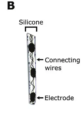
Figure 1. The clinical-grade electrode array used in the model, shown with its connecting wires prior to gel casting. Source: de Rijk et al. Tissue-Engineered Cochlear Fibrosis Model Links Complex Impedance to Fibrosis Formation for Cochlear Implant Patients. Advanced Healthcare Materials. 2023.
All three parts were modeled in Autodesk Fusion 360 and 3D printed at 30 µm XY resolution, with a tight enough tolerance to keep a multi-electrode array reliably centered inside a few millimeters of gel. The fibrin gel itself was made by mixing a fibrinogen stock solution with thrombin and calcium chloride directly around the electrode array, then left to react for two hours at 37°C before the whole construct was submerged in culture media inside a conical bioreactor. A contractile culture medium supplemented with TGF-β1 encouraged the seeded fibroblasts to behave more like the scar-forming cells seen in real fibrosis. From there, the team tracked each construct with electrochemical impedance spectroscopy (EIS) and full voltage waveform recordings at six time points across 14 days, alongside microscope imaging to watch the gel visibly contract around the array, comparing electrically stimulated constructs against unstimulated controls throughout.
Results
Over the 14-day culture period, the fibrin constructs contracted substantially around the electrode array, shrinking by an average of 60% ± 35% relative to their starting length. Histology confirmed this was not just a visual effect: denser cell packing and oriented collagen bundles had formed around the array, consistent with real fibrous tissue formation. This contraction ramped up sharply between days 7 and 9, the same window in which the model’s electrical readings began to shift.
Electrochemical impedance spectroscopy revealed a consistent pattern over time: resistance in the bulk of the construct rose significantly while its capacitance dropped, changes best explained by fibroblasts building up and reorganizing extracellular matrix around the electrode array rather than anything happening right at the electrode surface. Voltage waveform readings, the kind of signal a cochlear implant can already record without extra hardware, showed the same trend, giving the team a way to translate this lab finding into something clinically measurable.
From these waveforms, the team proposed a new marker called the second phase peak ratio (SPPR), calculated from a single existing stimulation pulse with no additional equipment required. When tested in 4 recently implanted cochlear implant patients over 5 months of follow-up, SPPR rose significantly between the two postoperative check-ins, tracking the same pattern seen in the lab model. Notably, SPPR shifted earlier than the standard contact impedance measurement already used in clinics today, suggesting it could flag fibrosis formation sooner and open a wider window for treatment intervention.
“…creating opportunities for earlier treatment intervention to improve cochlear implant efficacy.”
— de Rijk et al., Advanced Healthcare Materials, 2023
Products Used In This Study

Questions on how our 3D printing system can work for your research? Connect with one of our specialists.
More Research Article Summaries
McGill University researchers build a 3D printed double-sided mold system to shape and merge brain organoids on a chip
University of Hawaiʻi researchers 3D print wearable "sweatainer" devices with true microscale internal channels
Want to read more article summaries?
We summarize the latest microfluidics and 3D printing research.
Curated from peer-reviewed publications featuring the CADworks3D system.