
Research Article Summary
5 Minute Read
Ohio State researchers build a tumour-on-a-chip device using a 3D printed resin mold
Title
Characterization of a hybrid hydrogel for studying lymphocyte infiltration and migration in a 3-dimensional in vitro tumour stroma device.
Authors
Marco A Rodriguez, Saeed Derakhshesh, Mejalaa Mega Jayaseelan, et al.
Journal
Biofabrication, 18 (2026) 025006
DOI Link
Summary
Researchers at The Ohio State University designed a multi-layer microfluidic device for studying how immune cells move into solid tumours, casting the device from a 3D-printed resin master mold and pairing it with a tunable hybrid hydrogel. Rodriguez et al. show that the printed-mold route gives a fast, repeatable way to produce soft silicone devices with the fine internal channels these tissue models need.

Summary Author
This page was prepared by CADworks3D to summarize and highlight a peer-reviewed research article authored by independent researchers utilizing the CADworks3D system.
Title
Characterization of a hybrid hydrogel for studying lymphocyte infiltration and migration in a 3-dimensional in vitro tumour stroma device.
Authors
Marco A Rodriguez, Saeed Derakhshesh, Mejalaa Mega Jayaseelan, et al.
Journal
Biofabrication, 18 (2026) 025006
DOI Link
Key Results at a Glance
4 layers
Stacked device build
Top reservoir, top gel channel, bottom gel channel, bottom reservoir cast as one part
~15 µl
Tiny gel chambers
Each hydrogel channel holds about 15 µl, sized for microscope imaging
2 sec
Fast gel cure
UV crosslinking of the hydrogel finishes in roughly two seconds
~20 µm
Fine internal pores
The printed-mold device pairs with a hydrogel averaging about 20 µm pores
Objective
Solid tumours wrap themselves in a dense supporting tissue called the stroma. This stroma behaves like a wall. It slows down or blocks the immune cells, mainly T cells, that are supposed to find and kill cancer cells. Because of this wall, many immunotherapies that work well against blood cancers or in a dish fall short against real solid tumours. The drug may be doing its job, but the immune cells never physically reach the tumour to act. To understand where this breaks down, researchers need 3D models that let them actually watch immune cells try to push through tumour tissue, rather than just measuring the end result.
The usual lab tools each fall short in a different way. A flat dish of cells is too simple: it is only two dimensions, so it cannot capture how cells move up, down, and through a real tissue. Mouse and other animal models are more complete, but they swap in non-human cells and signalling pathways that do not always match what happens in a person, which makes the results harder to translate to patients. A stronger option sits in between: a small engineered device, built from human cells and a gel that mimics tumour tissue, where immune-cell movement can be imaged directly under a microscope.
The gap this paper addresses is a practical, fabrication-focused one. To build that kind of device, you need stacked tissue compartments that keep different cell types separate, and channels small enough to fit under a microscope objective. Just as important, you need to make the device the same way every time, so that any difference you see in cell movement comes from the biology and not from the device varying from run to run. The team’s answer was to design the device in CAD, 3D print a resin master mold, and cast the working device from that mold, then load it with a custom hydrogel tuned to let immune cells move through it.
Methodology and Design
The device is built around a 3D-printed mold that acts as a casting template. The mold is designed in CAD software and printed on a resin printer, then used to cast PDMS into the shape of the finished channels. Once cured and peeled away, the PDMS is bonded together into a stacked, multi-chamber structure and loaded with a tunable hydrogel matrix and cells to form the completed device.
The build breaks down into four core components, each covered in more detail below, including the working hydrogel: a gelatin, collagen, and PEG blend the authors call GCP:
Tumour-on-a-chip device
3D printed resin mold
GCP hybrid hydrogel
Stacked gel chambers
The finished platform is built for imaging immune cell behavior within a 3D tumor microenvironment. A silicone cover was pressed over the gel ports to seal the device against leaks, and growth media was added to the top and bottom reservoirs. The following day, immune cells were added to the top reservoir, and the team imaged how far those cells migrated through the gel over a six day window, comparing conditions with and without fibroblasts present.
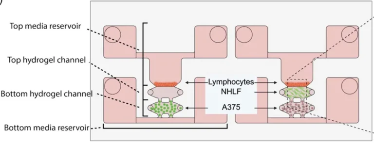
Figure 1. Cross-section of the finished device. Labels on the left mark the four stacked compartments, from the top media reservoir down to the bottom media reservoir. The central arrows show where each cell type sits: lymphocytes (immune cells) and NHLF (fibroblasts) in the top gel layer, and A375 (melanoma cells) in the bottom gel layer. Source: Rodriguez et al. Biofabrication. 2026.
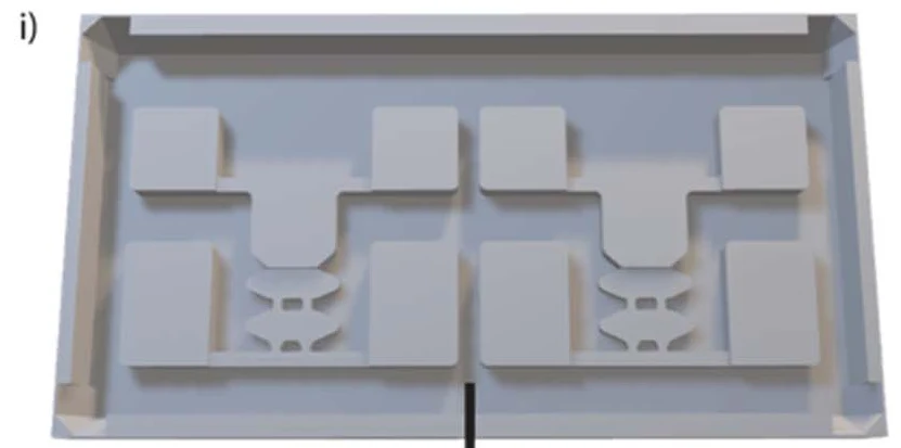
The mold that shapes the device started as a CAD drawing. The team designed a custom mold in AutoCAD, exported the design as STL files, and 3D printed the master mold on a resin printer using a mold-specific resin. This step is worth pausing on: the printed part is not the device itself. It is a reusable template, and its raised features map out every channel, port, and reservoir that will end up in the final device. Printing the mold rather than machining it means the geometry can be redesigned and reprinted quickly, and each print carries the same fine detail.
A short silane treatment coated the inside of the channels so the hydrogel would grip the walls instead of pulling away and leaking. Melanoma cells were mixed into the hydrogel, pipetted into the bottom gel channel, and UV cured in place in about two seconds. Fibroblasts, or a cell-free gel for control runs, went into the top channel and were cured the same way.
Liquid silicone, a material called PDMS, was poured over the printed mold and cured. As it set, it picked up the mold’s shape in negative, so the mold’s raised features became hollow channels in the silicone. Once peeled off, inlet and outlet holes were punched into the silicone using a biopsy punch, and the part was bonded to a glass coverslip using plasma treatment, which makes the two surfaces stick permanently. With the device built, cells were loaded layer by layer into the resulting stacked chambers.
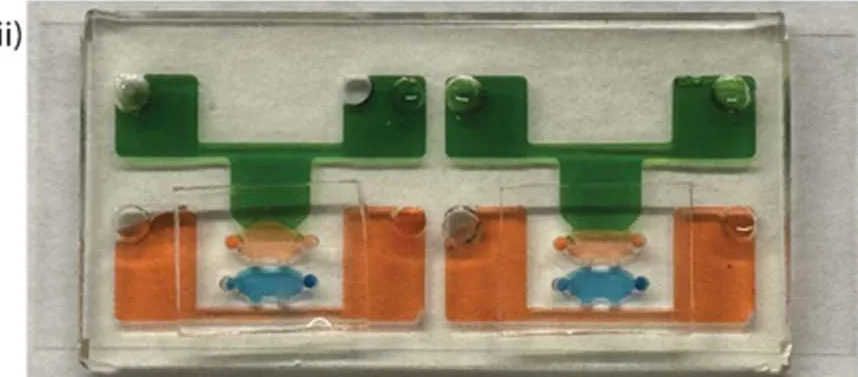
Figure 2. Two finished devices cast in silicone from the printed mold, with channels filled with coloured dye to show the stacked fluid paths. Source: Rodriguez et al. Biofabrication. 2026.
Results
The printed-mold approach delivered a device with exactly the stacked architecture the model needs: a four-layer build, with two media reservoirs sandwiching two hydrogel channels. Each gel chamber holds only about 15 µl, which is small enough to sit cleanly within the microscope’s field of view. Because the channels were defined by the printed master rather than shaped by hand, every cast device carried the same internal geometry. That repeatability matters when the whole point is to compare cell movement across many devices and many runs.
The fast 2-second UV cure was what made the layer-by-layer build practical. Each gel layer could be loaded and set almost instantly, so the cells already placed in the lower channel were not disturbed while the next layer went in. This is a direct benefit of pairing the printed device with a quick-curing gel, and it keeps the separate tissue layers cleanly compartmentalized.
When the team looked inside the cured hydrogel using electron microscopy, the device’s working gel, referred to as GCP, showed an open, porous surface that the comparison gels did not have. Its average pore size landed near 20 µm, smaller and more uniform than the alternatives tested. The authors link this surface porosity directly to the gel’s ability to let immune cells enter the tissue in the first place, which is the behaviour the whole device is built to capture.
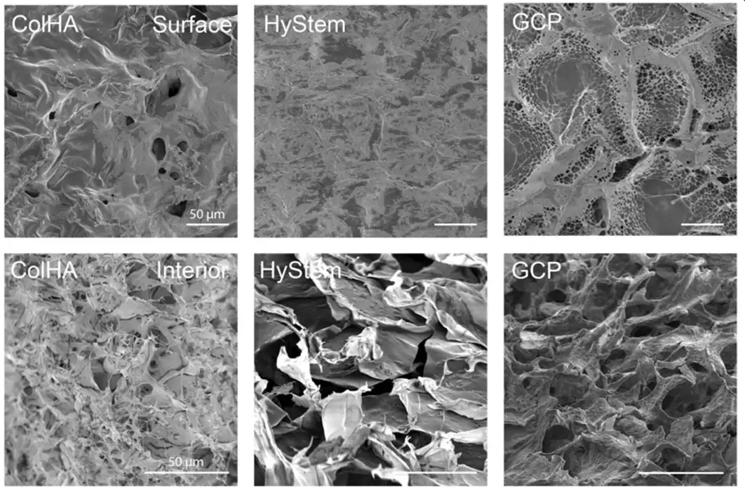
Figure 3. Electron microscope images comparing the surface and interior pore structure of three hydrogels. The GCP gel (right) shows the open, porous surface the device relies on. Source: Rodriguez et al. Biofabrication. 2026.
“A custom PDMS mold was designed with AutoCAD and STL files were transferred to a resin printer to manufacture using mold resin.“
— Rodriguez et al., Biofabrication, 2026
Products Used In This Study

Questions on how our 3D printing system can work for your research? Connect with one of our specialists.
More Research Article Summaries
McGill University researchers build a 3D printed double-sided mold system to shape and merge brain organoids on a chip
University of Hawaiʻi researchers 3D print wearable "sweatainer" devices with true microscale internal channels
Want to read more article summaries?
We summarize the latest microfluidics and 3D printing research.
Curated from peer-reviewed publications featuring the CADworks3D system.