
Research Article Summary
5 Minute Read
NC State and UNC researchers fabricate H-channel microfluidic chips from 3D printed molds to profile fibrin behavior
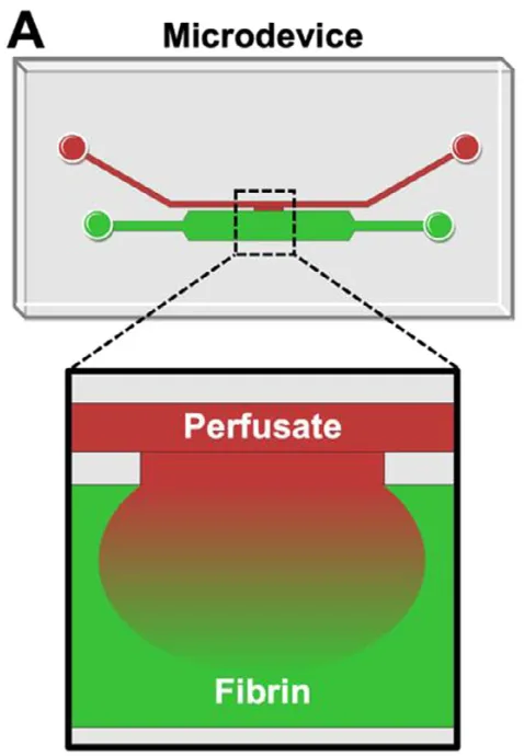
Title
Microfluidic Platform for Multiparametric Profiling of Fibrin Permeability, Fibrinolysis, and Cell Invasion
Authors
Halston Deal, Elizabeth M. Byrnes, Sanika Pandit, et al.
Journal
Advanced Functional Materials, (2026)
DOI Link
Summary
Researchers built a single-channel, H-junction microfluidic device using 3D printed molds that lets them measure fibrin permeability, clot breakdown, cell invasion, and clot growth all within the same tiny hydrogel sample. By casting PDMS chips from resin-printed molds rather than relying on traditional photolithography, the team (Deal et al.) created a fast, repeatable fabrication route for studying how blood clots form and dissolve.
Summary Author
This page was prepared by CADworks3D to summarize and highlight a peer-reviewed research article authored by independent researchers utilizing the CADworks3D system.
Title
Microfluidic Platform for Multiparametric Profiling of Fibrin Permeability, Fibrinolysis, and Cell Invasion
Authors
Halston Deal, Elizabeth M. Byrnes, Sanika Pandit, et al.
Journal
Advanced Functional Materials, (2026)
DOI Link
Key Results at a Glance
50 µm
Print Slicing Resolution
Layer height used to print the device molds
~3 µL
Analysis Volume
Gel region needed per quantitative measurement
4-in-1
Multiparametric Chip
Permeability, lysis, cell invasion, and clot growth in one device
240 / 130 µm
Channel Heights
Flow/fibrin channels at 240 µm, interfacial channel at 130 µm
Objective
Blood clotting depends on fibrin, the insoluble mesh that forms when the soluble protein fibrinogen is converted by thrombin. Fibrin does two very different jobs depending on context. In a healing wound, it acts as a temporary scaffold that holds cells and growth factors in place while tissue rebuilds. In disease, the same material becomes a problem, showing up as the bulk of dangerous clots in deep vein thrombosis, atherosclerotic plaques, and several neurological conditions. Because fibrin sits at the center of both repair and pathology, researchers need tools that can describe how it transports fluid, how quickly it breaks down, and how readily cells move through it.
The challenge is that studying these properties has traditionally meant juggling several separate, equipment-heavy experiments. Permeability testing alone has historically required bulky assemblies of pipettes, tubes, beakers, and ring stands, and each of these setups consumes relatively large sample volumes. That matters a great deal for fibrin research, because some of the most scientifically interesting fibrinogen, such as fetal or disease-specific samples, is difficult and expensive to obtain. Running permeability, breakdown, and cell migration as three disconnected assays also introduces variability, making it harder to compare results and contributing to the broader reproducibility problem across biomedical research.
A second gap is fabrication. Microfluidic devices are usually made through photolithography, a multi-step cleanroom process that is slow, costly, and not easy to iterate on. For a lab that wants to test several channel geometries quickly, photolithography becomes a bottleneck. The researchers set out to address both gaps at once: consolidate four fibrin measurements into a single standardized chip, and replace the cleanroom mold-making step with a faster route built around 3D printing.
The device they designed uses an H-shaped channel arrangement. Two parallel channels (a “flow” channel and a “fibrin” channel) are joined by a narrow interfacial channel, and fibrin is held in place at this junction. By moving fluorescent dyes, plasmin, cells, or whole blood through the flow channel and watching how signals change at the interface under a microscope, the team can run every assay using the same imaging approach. The lateral, side-by-side layout was chosen deliberately because it is easier to track under a microscope and leaves room for future on-glass electronic sensors.
Methodology and Design
The fabrication workflow used in the study relies on resin 3D printing paired with soft lithography. Channel geometries are designed in CAD software and exported as printable files, then 3D printed as molds. The post-processing sequence follows the mold through an isopropanol rinse, UV post-cure, PDMS casting, PDMS removal, and plasma bonding to seal the finished device.
All four applications ran on the same H-channel architecture, which is the point of the design: one fabrication workflow, one imaging setup, four readouts.To benchmark the resin, four devices spanning both liquid-handling and biological applications were built and tested:
Permeability Chip
Fibrinolysis Chip
Cell Invasion Chip
Clot Growth Chip
The measurement principle was kept identical across assays so the chip would be easy to adopt. In every case, something fluorescent moves through or along the fibrin gel and a confocal microscope tracks how the signal changes over time. For permeability, fluorescent dextran or rhodamine dye diffuses into the gel. For breakdown, the enzyme plasmin is perfused and the loss of fibrin signal is timed. For cell work, dyed fibroblasts are seeded and their advance into the gel is imaged over five to seven days. For clot growth, recalcified whole blood spiked with fluorescent fibrinogen is flowed past a preformed gel. Because the geometry is fixed and the imaging is shared, results from one assay can be compared directly against the others.
Channel geometries were drawn in AutoCAD and exported as STL files at a facet resolution of 10, the format a resin printer reads. The molds were then printed at 50 µm layer slicing. The team used two different resins depending on the experiment. A dedicated mold resin was used for the validation and multiplexing devices, which featured 500 µm wide flow channels with a 200 µm gap to the fibrin channel. A clear resin was used for the adult-versus-fetal comparison devices, which were built with finer features, a 300 µm wide flow channel and an 85 µm gap.
Post-processing differed by resin and is where most of the hands-on care happened. Prints were soaked in isopropyl alcohol to clear uncured resin, five minutes for clear resin and twenty minutes for the mold resin, then sprayed with acetone followed by IPA, dried with compressed air, and UV cured. Cure times again diverged sharply: three minutes for clear resin versus 100 minutes for the mold resin. The clear resin molds received one extra step, a roughly 3 µm parylene C coating, which improves the mold surface before casting.
With the molds finished, the team moved to soft lithography. PDMS, a flexible silicone, was mixed at a standard 10:1 base-to-crosslinker ratio, poured over the printed molds, degassed to remove bubbles, and baked at 70 to 80 °C for at least two hours. The cured PDMS was peeled off, cut into individual channel segments, and inlet/outlet ports were punched with biopsy punches (1.5 mm for cell-free experiments, 4 mm where cells needed room to be seeded). Each PDMS segment was then plasma bonded to a glass coverslip or microscope slide to seal the channels into a finished device. Devices destined for permeability and breakdown experiments were baked at 80 °C for two to three days afterward to restore the surface properties needed for clean flow.
Results
The fabrication approach worked, and the chip resolved clear, quantitative differences across every fibrin condition tested. Crucially, the entire quantitative analysis happened in a gel region of only ~3 µL, a major reduction from well-plate methods that required 200 µL of clot solution per replicate.
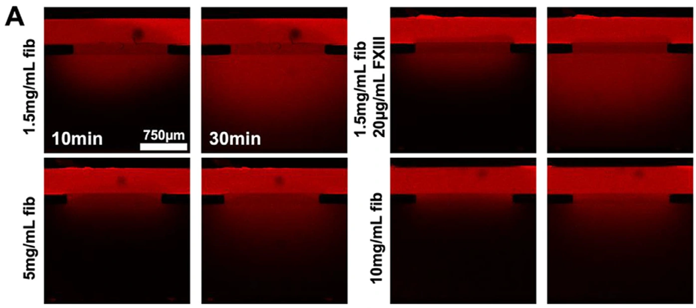
Figure 1. Confocal images of dye permeating fibrin gels of increasing concentration, showing how denser networks slow solute transport. (Scale bar 750 µm.) Source: Deal et al. Adv Funct Mater. 2026.
Increasing fibrinogen concentration produced denser networks and measurably lower transport. Permeability fell across a 3.7-fold span, dropping from 7.35 × 10⁻⁵ cm²·s⁻¹ in the loosest 1.5 mg·mL⁻¹ gels to 1.98 × 10⁻⁵ cm²·s⁻¹ in the densest 10 mg·mL⁻¹ gels. Confocal imaging confirmed the cause, with fiber pixel density climbing from 17.4% to 35.1% as concentration rose.
Clot breakdown followed the inverse trend, as expected for tighter networks. Across the conditions tested, lysis rate shifted up to ninefold, with looser gels dissolving fastest. The chip also captured a more subtle biological result: the crosslinking factor FXIII had only a modest effect on adult fibrin but significantly increased the breakdown resistance of fetal fibrin, which otherwise lysed most rapidly at 11.4% per minute versus 4.03% per minute for adult fibrin in the first six minutes.
Cell invasion tracked the same density story. Denser fibrin suppressed fibroblast penetration by up to 64%. In the loosest gels, cells kept a 3D migration morphology and pushed past 750 µm from the origin, while in denser gels they shifted toward shallow, surface-level 2D movement. Comparing sources, fetal fibrin (lower density, higher permeability) consistently allowed faster and deeper infiltration than adult fibrin. Finally, the whole-blood demonstration confirmed the chip can model clot growth, with recalcified blood producing more than double the fibrin signal of untreated blood at the gel boundary by 30 minutes.
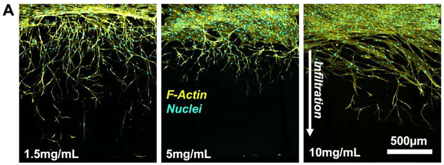
Figure 2. Fibroblasts infiltrating gels of three densities. Looser gels permit deep 3D migration; denser gels push cells toward shallow movement. (Scale bar 500 µm.) Source: Deal et al. Adv Funct Mater. 2026.
“Molds for our devices can also be 3D printed, avoiding more laborious photolithography.”
— Deal et al., Advanced Functional Materials (2026)
Products Used In This Study


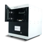
Questions on how our 3D printing system can work for your research? Connect with one of our specialists.
More Research Article Summaries
McGill University researchers build a 3D printed double-sided mold system to shape and merge brain organoids on a chip
University of Hawaiʻi researchers 3D print wearable "sweatainer" devices with true microscale internal channels
Want to read more article summaries?
We summarize the latest microfluidics and 3D printing research.
Curated from peer-reviewed publications featuring the CADworks3D system.