
Research Article Summary
5 Minute Read
LSU researchers spin microscopic spheres inside a 3D printed resin chamber to lay groundwork for handling zebrafish eggs
Title
A 3D-printed microdevice for dielectrophoretic torque-driven rotation of dielectric microspheres to support development of manipulation systems for zebrafish eggs
Authors
Seyedmajid Hosseini, Mohsen Norouzi, Jose Cibelli, et al.
Journal
Journal of Micromechanics and Microengineering, 35 (2025) 125005
DOI Link
Summary
Researchers fabricated a resin 3D printed microdevice that uses a rotating electric field to spin 700 µm dielectric microspheres without ever touching them, a step toward non-contact handling of zebrafish eggs in cloning workflows (Hosseini et al.).

Summary Author
This page was prepared by CADworks3D to summarize and highlight a peer-reviewed research article authored by independent researchers utilizing the CADworks3D system.
Title
A 3D-printed microdevice for dielectrophoretic torque-driven rotation of dielectric microspheres to support development of manipulation systems for zebrafish eggs
Authors
Seyedmajid Hosseini, Mohsen Norouzi, Jose Cibelli, et al.
Journal
Journal of Micromechanics and Microengineering, 35 (2025) 125005
DOI Link
Key Results at a Glance
~20 min
Print Time Per Device
Each microdevice prints in about twenty minutes
< $1
Resin Cost Per Device
Material for one device costs under a dollar
~100x
Cheaper Than Phtolighography
Cost cut by roughly two orders of magnitude
850 µm
Printed Chamber Diameter
Sized to cradle a single egg-scale object
Objective
Handling something as small and delicate as a zebrafish egg is one of the quiet bottlenecks in reproductive biology. Eggs in the 700 to 800 µm range have to be trapped, held still, and rotated so that a single narrow opening called the micropyle lines up with an injection needle. In a technique called somatic cell nuclear transfer, where the nucleus of one cell is moved into an egg whose own nucleus has been removed, that alignment step is critical. Today it is usually done by hand under a microscope using fine glass pipettes or mechanical micromanipulators. That work is slow, depends heavily on the skill of the operator, and takes a long time to learn.
There is a cleaner way to move tiny objects: use electric fields instead of physical probes. When an object that conducts poorly is placed in a rotating field, the field induces a temporary charge separation inside it, and the lag between the field and that induced charge produces a twisting force, or torque. The object spins in place without anything touching it. This idea, electrorotation, is a close cousin of dielectrophoresis, and both are attractive because they are label-free, contactless, and easy to tune by changing the frequency or strength of the field.
The catch is how these field-generating devices have traditionally been built. Most prior electrorotation work used electrodes patterned in a cleanroom through photolithography, a process that is expensive, slow, and out of reach for many labs. It also tended to focus on much smaller targets, single cells or particles only a few µm across, rather than the comparatively giant 700 µm objects that an egg represents. Very little work had tried to rotate objects at egg scale, and the few attempts still leaned on costly conventional microfabrication.
This is the gap the study addresses. The authors set out to build the first resin-printed electrorotation microdevice designed specifically for large, egg-scale objects, using a desktop resin printer instead of a cleanroom. The goal was a platform that is fast to make, cheap enough to iterate on freely, and easy for other labs to copy and modify. Their specific aims were to design and print the device at high resolution, model how field frequency and material properties should drive rotation, simulate the behavior before building, and then validate the printed device experimentally with real microspheres.
Methodology and Design
The device began as a CAD model designed in Fusion 360, which let the team control geometry and electrode placement precisely before anything was printed. The body was then produced directly on a desktop resin printer at high resolution, a deliberate move away from photolithography, with copper wire electrodes slid into printed guide channels to complete the assembly.
The fabrication process used in the study utilizes digital light processing (DLP) 3D printing. Device geometries are designed in CAD software (e.g., SolidWorks), then sliced and 3D printed. After printing, the post-processing workflow follows the mold undergoing: an isopropanol rinse → UV post-cure → PDMS casting → PDMS removal → plasma-bonding. During the PDMS casting stage, Sylgard 184 PDMS (10:1 base-to-curing-agent) was degassed for 15 minutes, poured directly onto the printed mold with no silanization or solvent treatment, oven-cured, peeled, punched, and plasma-bonded to glass or PDMS.
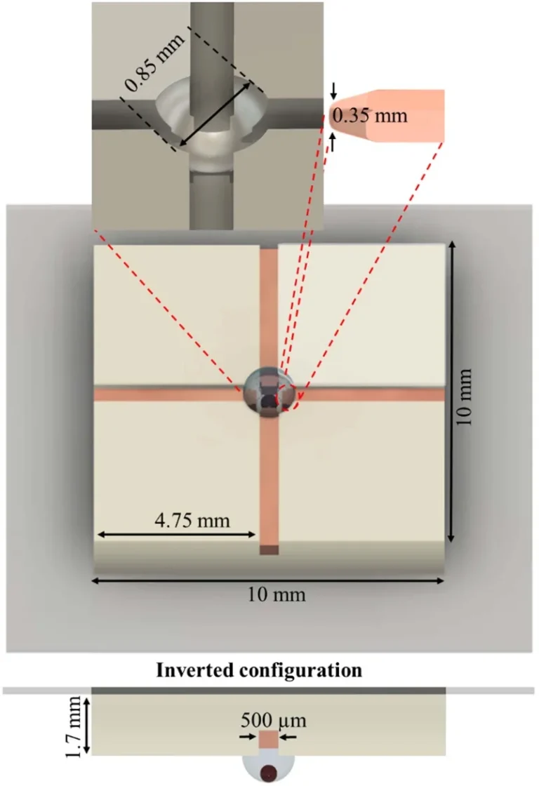
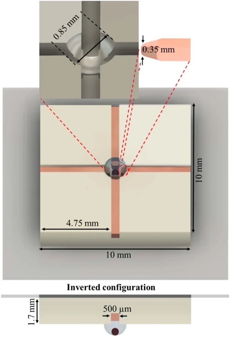
Figure 1. CAD schematic of the resin printed device, showing the quadrupole copper electrodes, the 850 µm chamber, and the inverted hanging-droplet configuration with key dimensions. Source: Hosseini et al. A 3D-printed microdevice for dielectrophoretic torque-driven rotation of dielectric microspheres. J. Micromech. Microeng. 2025.
To achieve controlled rotation of microsphere and egg-scale samples, the printed platform combines three core components:
Electrorotation Microdevices (ERM)
Semispherical Printed Chamber
Semispherical Printed Chamber
Each unit printed in about twenty minutes and used less than a dollar of resin, which made it cheap to print many versions and refine the design quickly. No cleanroom and no special alignment steps were needed, so assembly stayed simple enough to repeat across labs.
At the center of the printed body sits a semispherical chamber 850 µm wide, sized to hold a single egg-scale object such as a zebrafish egg, which typically runs 700 to 800 µm across. This chamber was printed in a clear microfluidic resin so the object inside could be watched and aligned under a microscope during testing. Around the chamber, four copper wire electrodes with square 500 µm cross-sections were slid into printed guide channels matched to their dimensions. A small printed shoulder inside each channel acted as a stop, halting each electrode at a fixed depth so it could not be pushed in too far. That detail kept all four electrodes positioned consistently relative to the chamber center, which matters because the field has to stay symmetric. The four electrodes were arranged in a square, the quadrupole layout that produces a rotating field, and the printed base doubled as the fixture that connected them to standard clips.
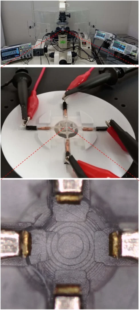
Figure 2. The full experimental setup, with the printed microdevice wired to signal generators, an oscilloscope, and a microscope. The close-up shows the four electrodes meeting at the central semispherical well. Source: Hosseini et al. A 3D-printed microdevice for dielectrophoretic torque-driven rotation of dielectric microspheres. J. Micromech. Microeng. 2025.
To make the field actually rotate, the four electrodes were each driven by an alternating signal shifted in phase by 90 degrees relative to its neighbor, produced by two synchronized signal generators and checked on an oscilloscope. A single 700 µm polystyrene bead was pipetted into the chamber in a saline buffer (DPBS) chosen to be gentle on biological samples. Early on, friction between the bead and the chamber floor stopped it from turning, so the team simply flipped the device over. Inverted, the bead hung from the chamber ceiling inside a hanging droplet, which cut its contact with the surface and let it spin freely. Rotation was filmed through an inverted microscope and measured frame by frame. Before building, the whole system was also simulated in COMSOL to confirm the field would form correctly and to fine-tune the electrode geometry without reprinting. The electrode tips were rounded rather than sharpened, since blunt tips spread the field more evenly around a large object and avoid the localized heating that sharp tips can cause.
Results
The printing approach delivered on its central promise. Each device took roughly twenty minutes to print and cost under a dollar in resin, cutting both labor and material cost by about two orders of magnitude and fabrication time by at least one order of magnitude compared with photolithography. The printed body carried a working quadrupole electrode layout around its 850 µm chamber, and signal testing confirmed all four electrodes reliably received their phase-shifted inputs. Assembly needed no cleanroom, and the open, shareable design means other labs can reproduce it directly.
The device worked as a rotation platform. A single 700 µm polystyrene bead rotated steadily clockwise once the field was applied, and the inverted, hanging-droplet arrangement was the key fix that turned stalled beads into smooth, continuous spinning. The strongest rotation reached about 8.1 degrees per second at 900 kHz and 16 Vp-p. The simulations and the underlying theory predicted the rotation should follow a single-peak response that rises and then falls with frequency, and the experiments matched that shape, even though the real-world peak landed lower than the modeled one, a gap the authors attribute to real device factors like electrode edges, droplet curvature, and heating.
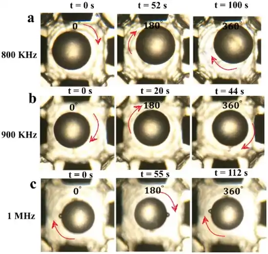
Figure 3. Time-lapse microscope views of a 700 µm bead rotating clockwise inside the printed chamber at 800 kHz, 900 kHz, and 1 MHz. Red arrows mark the direction of rotation. Source: Hosseini et al. A 3D-printed microdevice for dielectrophoretic torque-driven rotation of dielectric microspheres. J. Micromech. Microeng. 2025.
There were practical limits worth noting. Below about 16 Vp-p the bead would not turn at all, while pushing past it tended to form bubbles that destabilized the field, so 16 Vp-p sat in a usable window. Rotation also failed entirely in deionized water, which lacks the dissolved ions needed to generate torque on an object this large, confirming that the gentle saline buffer was the right medium for future biological use. Together these results establish the printed device as a low-cost, reproducible foundation for eventually rotating and aligning real zebrafish eggs.
“Resin 3D printing turned a process that once demanded a cleanroom into a twenty-minute, sub-dollar print that any lab can copy and modify.“
— Hosseini et al., Journal of Micromechanics and Microengineering, 2025
Products Used In This Study

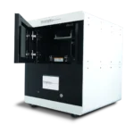
Questions on how our 3D printing system can work for your research? Connect with one of our specialists.
More Research Article Summaries
McGill University researchers build a 3D printed double-sided mold system to shape and merge brain organoids on a chip
University of Hawaiʻi researchers 3D print wearable "sweatainer" devices with true microscale internal channels
Want to read more article summaries?
We summarize the latest microfluidics and 3D printing research.
Curated from peer-reviewed publications featuring the CADworks3D system.