
Research Article Summary
5 Minute Read
Illinois and Biohub Chicago researchers build a reusable, modular microfluidic platform that cuts device prep from days to about two hours
Title
Rapid and reusable high-throughput microfluidics through modular assembly
Authors
Linh T.P. Le, Omkar Hegde, Wei-Huan Wu, et al.
Journal
BioRxiv (preprint, posted January 13, 2026; not yet peer-reviewed)
DOI Link
Summary
Researchers led by Le et al. developed a modular high-throughput microfluidic platform that can be disassembled, cleaned, and reused instead of being thrown away after a single experiment, dropping setup time from several days to roughly two hours while preserving dense microvalve-driven fluid control.
Summary Author
This page was prepared by CADworks3D to summarize and highlight a peer-reviewed research article authored by independent researchers utilizing the CADworks3D system.
Title
Rapid and reusable high-throughput microfluidics through modular assembly
Authors
Linh T.P. Le, Omkar Hegde, Wei-Huan Wu, et al.
Journal
BioRxiv (preprint, posted January 13, 2026; not yet peer-reviewed)
DOI Link
Key Results at a Glance
~2 hours
Reset time between experiments
Down from the conventional 3 to 4 day fabrication cycle.
~50 µm
Bonded PDMS membrane thickness
Permanently sealed beneath each control hole to absorb high pressure.
1 mm
Deep-well chamber depth
Cast from a 3D printed master for 3D organoid culture.
160
Individually controllable chambers
Roughly double the count of the team’s previous chip design.
Objective
Microfluidic devices let researchers move and control tiny volumes of fluid, often at the scale of a few tens of µm, which is close to the size of a single biological cell. This makes them powerful tools for experiments like single-cell analysis, drug screening, and growing miniature tissues. To get the most out of a single chip, designers pack in as many channels and chambers as possible. Densely packed features let many independent experiments run side by side on one device, dramatically increasing throughput.

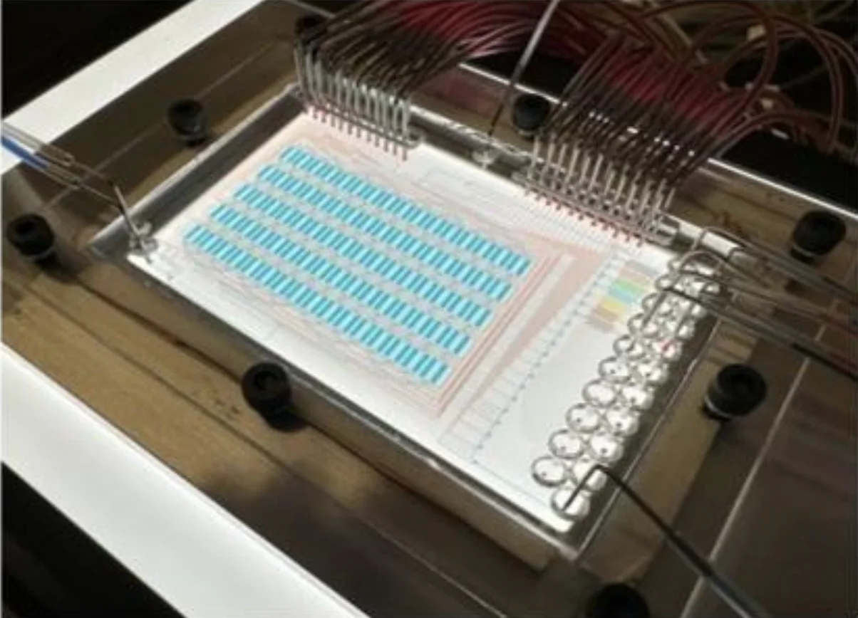
Figure 1. An exploded view of the modular platform shows how the microfluidic chip, chamber layer, and glass substrate stack inside a custom aluminum tray, alongside a workflow comparing the reusable modular route against conventional single-use fabrication. Source: Le et al. bioRxiv. 2026.
But that density comes at a cost. Narrow channels are easy to clog, dust or debris can block flow or cause neighboring channels to leak into each other, and the thin PDMS layers that make up the device can shift slightly during assembly and ruin the alignment. Building these multilayer chips requires high-precision molds, careful layer-by-layer bonding, and a fabrication process that can take three to four days. A single mistake at any step means starting over.
The bigger problem is that most of these devices are effectively single-use. Once a chip is clogged or contaminated, the microscopic channels are nearly impossible to clean. And because devices are usually designed for one specific experiment, even a small change in the research question often means redesigning and refabricating the whole thing. The result is an expensive, slow, failure-prone workflow that keeps high-throughput microfluidics locked inside specialized, well-funded engineering labs.
The gap this paper addresses is straightforward to state but hard to solve: how do you keep all the benefits of a dense, automated, multilayer microfluidic chip while making it reusable, faster to prepare, and adaptable to different experiments? The authors’ answer is a modular design that separates the complicated, expensive, reusable part of the device from the cheap, disposable part that actually touches the cells.
Methodology and Design
The platform is built around two separate modules: a reusable fluid-control module that houses the plumbing, and a replaceable substrate, the surface cells actually grow on. The control module consists of two PDMS microchannel layers, with pressurized control channels on top flexing a thin membrane down to pinch the fluid channels below shut, acting as microvalves. The substrate is the part that gets contaminated with each experiment, so it’s designed to be cheap, mass-produced in batches, and swapped out for every run. Rather than bonding it permanently, the team used reversible bonding so the whole stack can be taken apart, cleaned, and reassembled with a fresh substrate each time.
This modular design was applied across three chip formats to demonstrate its versatility:
Thin-Substrate Chip
Deep-Well Chip
Macrophage Assay
The substrate comes in interchangeable formats depending on the experiment. For high-resolution 2D imaging of single cells, a glass slide is spin-coated with a thin hybrid adhesive polymer, a 40:1 blend of a soft silicone adhesive and PDMS, creating a reversible seal that’s clean enough for microscopy and sterile for cell culture. For 3D organoid work, the team used a deep-well substrate instead, cast from a 3D printed mold to form chambers about 1 mm deep. Loading cells into these open wells before assembly, much like seeding a well plate, keeps the microscopic channels clean and avoids pushing cells or thick media through narrow passages. The same modular platform also supports a macrophage assay for studying immune signaling.
The control module’s two PDMS layers are separated by a thin membrane of about 20 µm. Because the valves this membrane forms often need more than 10 psi to fully close, the two layers are permanently plasma-bonded together so they never come apart. To fabricate the module, the thick control layer is cast from PDMS, trimmed, and punched with access holes before it’s aligned and bonded to the thin, spin-coated fluid layer below. A key detail in the workflow is that these holes are punched before that bonding step, which leaves a roughly 50 µm PDMS membrane permanently bonded beneath each control hole. This cushions the more fragile substrate bond against the high pressures applied during operation.
For the deep-well substrate, additive manufacturing enters the workflow directly. Positive molds were designed in CAD, processed, and printed on a high-resolution resin 3D printer. The printed molds were rinsed in isopropanol, air-dried to clear their fine features, and post-cured under 405 nm UV light, then PDMS was cast against these printed masters to form the finished chambers.

Figure 2. Fabrication workflows for the reusable PDMS module and its two substrate types, including the deep-well substrate cast from a 3D printed master to create 1 mm chambers for 3D culture. Source: Le et al. Rapid and reusable high-throughput microfluidics through modular assembly. bioRxiv. 2026.
Between runs, the reusable module is flushed with water and air, peeled off the spent substrate, rinsed with pressurized water, optionally ultrasonicated and autoclaved, dried, and baked at 60°C for at least 30 minutes. After this, it’s ready to bond to a new substrate. The chip channels were also pre-coated with Pluronic or Teflon to reduce sticking, helping the same module run reliably across many experiments.
Results
The modular approach delivered its headline benefit cleanly: where conventional multilayer fabrication runs a 3 to 4-day cycle with failure risk stacking up at every step, swapping in a fresh substrate cut per-experiment setup to under 2 hours. Because the most failure-prone and labor-intensive component, the fluid-control module, is reused rather than rebuilt, the cost and time of each new experiment dropped dramatically.
Reversible bonding strength came down to the substrate coating. The hybrid adhesive polymer performed best, holding up to 4.2 to 4.5 psi before delaminating, while a 20:1 PDMS coating tolerated up to 3.2 psi, which is enough for most applications when negative pressure is also applied at the outlet. Plain uncoated glass performed worst. Critically, when the substrate was replaced with a fresh one for each run, burst pressure stayed consistent across repeated trials, confirming the module itself holds up to reuse. Reusing the same adhesive substrate, by contrast, dropped burst pressure to about 1 psi, since the soft adhesive layer tears during disassembly. This is why the team treats the substrate as disposable and the module as durable.
The redesigned chip layout also boosted throughput. By clustering control-line holes away from the fluidics and adding serpentine channels to balance flow, the team roughly doubled the chamber area (1.8 to 3.4 mm²) and increased the chamber count to 160 individually controllable chambers, up from 64 to 92 in earlier designs. Automated routing tests filled individual chambers in letter-shaped patterns with no cross-contamination between neighbors.
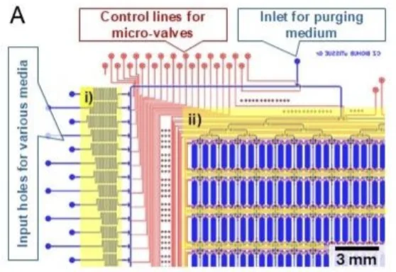
Figure 3. CAD layout of the chip's fluid channels (blue) and control channels (red), showing the serpentine paths that balance flow and the multiplexers that expand sample count. Source: Le et al. bioRxiv. 2026.
Finally, the platform held up under a demanding biological test. Mouse macrophages were exposed to changing doses of LPS while NF-κB signaling was tracked by fluorescence imaging. The device stayed leak-free and mechanically stable across repeated pressure cycles, and cells stayed healthy through a 12-hour perfusion experiment, confirming the hybrid adhesive interface is biocompatible. The team even reused the fluid-control module from one trial directly into the next, demonstrating reuse in a real experiment rather than just a benchtop test.
“This approach improves repeatability and experimental success while reducing preparation time from days to ~2 hours.“
— Le et al. bioRxiv. 2026.
Products Used In This Study
Questions on how our 3D printing system can work for your research? Connect with one of our specialists.
More Research Article Summaries
McGill University researchers build a 3D printed double-sided mold system to shape and merge brain organoids on a chip
University of Hawaiʻi researchers 3D print wearable "sweatainer" devices with true microscale internal channels
Want to read more article summaries?
We summarize the latest microfluidics and 3D printing research.
Curated from peer-reviewed publications featuring the CADworks3D system.