
SEASON 02 EPISODE 03
How does human biology change in space?
8th September, 2026
Hemdeep Patel, Robin Boshoven and Dr. Cassandra Juran
Podcast Summary
This episode of Big Ideas at Microscale features Dr. Cassandra Juran of Embry-Riddle Aeronautical University and NASA Ames Research Center, whose background spans aerospace engineering, tissue engineering, stem cell biology, and aerospace physiology. Her research explores how the human body responds to the unique physical forces of spaceflight, with a particular focus on mechanotransduction and the effects of microgravity on cellular and organ function.
Juran discusses how her team uses 3D-printed microfluidic devices and multi-organ tissue chips to study the effects of spaceflight on interconnected biological systems. By linking brain, liver, gut, lung, and bone marrow organoids, these organ-on-a-chip models provide a way to study space biology and microgravity without relying solely on animal models. The conversation also explores how 3D printing is enabling rapid prototyping of biomedical devices for space research, including tools designed for use aboard the International Space Station.
The episode looks toward the future of personalized space medicine, including the development of “astronaut-on-a-chip” systems that could help predict how individual astronauts will respond to deep-space hazards such as radiation and bone loss. Juran also shares how advances in biocompatible materials and 3D printing could accelerate research into human health during long-duration space missions.
Available On :



"We created that blood vessel layer at the bottom, and then we put the organoids above it. Using the three dimensionality and capability of 3D printing is something that is actually super powerful..."
Transcript
Transcript
Robin (00:00) Welcome back to Big Ideas at Microscale. We are really excited about today’s guest. We have Dr. Cassandra Juran, a stem cell and regenerative biologist specializing in mechanotransduction. She is Co-Director of Space Lab at Embry-Riddle Aeronautical University and an Associate Research Scientist with the Blue Marble Space Institute of Science at NASA Ames, in the Bone and Cell Signaling Lab. Welcome, Cassie. And joining me as always is my co-host Hemdeep.
Hemdeep (01:43) Hello everyone. I should mention that I did my undergraduate work in physics and astronomy, and NASA has always been a pinnacle of space exploration to me. But today I get to explore a side of NASA I don’t often see — the biology and astrobiology happening in orbit. I am so excited to talk with you, Cassandra. Thank you for taking time out of your busy day.
Cassandra (02:55) Thanks for the opportunity. I have listened to your podcast, so I was very excited to be a part of it.
Hemdeep (03:04) You have got two very impressive titles. Can you give us an idea of what each one entails and what else you are up to?
Cassandra (03:20) Sure. To understand my somewhat unusual titles, you have to understand me a little. I am not someone who knew what they wanted to do from age three and went straight for it. I was always very interested in spaceflight. I grew up here in Florida watching the shuttle launches. My dad was in the Air Force and later worked for the FAA, and my mom loved everything sci-fi. So I knew I wanted to be involved in space. I just did not quite know how.
I was actually terrible at biology in K through 12. I could not tell you what a bacterium was versus a plant. But I was strong in math and science, so I went into aerospace engineering at the University of Florida, graduated with honors, and was on track to work at United Launch Alliance, the company that handles rocket assembly and safety checks. Then the shuttle program ended and I lost the job before I even started.
That left me at a crossroads. I could have gone into aviation, Boeing, Piper, any of the major firms, but I decided to stay in school. I was working in control system theory at the time, specifically designing automated trajectories for returning spacecraft safely to Earth. One of those was the skip reentry trajectory that Artemis 1 used to come back from its trip around the moon. It is all control theory: figuring out how to guide a system through any kind of unpredictable environment and bring it home safely.
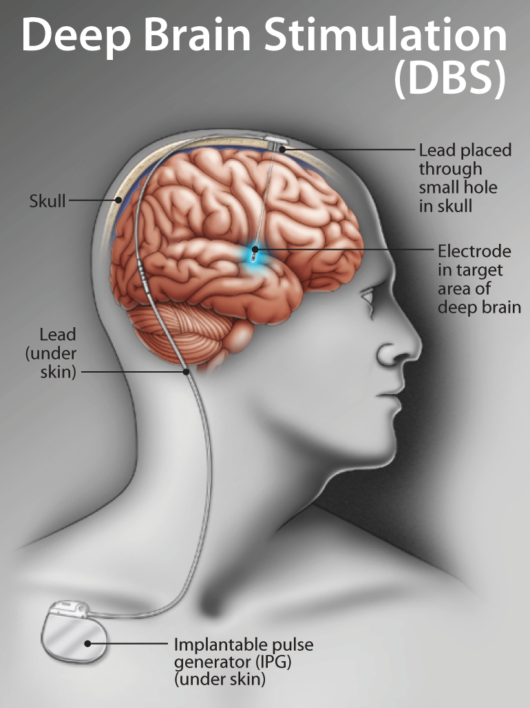
Cassandra (05:14) Because I was doing that work, a friend in the movement disorders clinic at the medical school asked if I would help him with a research project on Parkinson’s disease. He was studying deep brain stimulation, a technique where an electrical conductor is implanted in the brain and used like a pacemaker to normalize electrical activity and reduce tremors. At the time, only a handful of universities and clinics offered it, and patients were driving to the University of Florida from as far as Arkansas, twice a week, just to have their controllers manually tuned.
Knowing I wrote automated trajectory controllers, he asked: could you write an automated controller to manage biology? I had never thought about it, but I said I do not see why not. It is an electrical system that takes commands just like anything else. That was my first real entry into biology, entirely from an engineering perspective.
Hemdeep (07:42) So you literally approached the brain like a control system.
Cassandra (07:48) Essentially, yes. And that curiosity stayed with me. I still had a deep love of space, so for my PhD, still at the University of Florida, I moved into tissue engineering. My project focused on the most mechanically active joint in the human body: the temporomandibular joint, the small joint in your jaw. What drew me to it was that it is constantly moving, constantly under physical load, and the stem cells that live in that tissue are perpetually remodeling it in response. If you are isolated and barely talking for six months, the tissue adapts. If you are eating tough foods or talking constantly, it adapts differently. It was my first real biology project. Here are these squishy living things, and they are governed by the same dynamic mechanical forces I studied in engineering. Could I combine the two to treat disease in that joint?
Hemdeep (09:56) So you found a spot in the body that was naturally mechanical.
Cassandra (09:57) Exactly. And the logical next question was: where are we not mechanically loaded? Where do we stop experiencing gravity? That led me straight into space research. And that is where my titles finally make sense.
I am an assistant professor at Embry-Riddle in aerospace physiology, looking at how human biology changes in the extreme environments of aviation and space. Fighter pilots experience massive G-loading during maneuvers and their cardiovascular systems have to process enormous mechanical forces. Astronauts are in freefall, experiencing no gravitational acceleration, and are exposed to high doses of radiation, which is its own kind of cellular activator. So it was this long path from engineering, through biology, to trying to understand: how can I keep astronauts healthy in space, and how can space help us understand and treat disease on the ground?
My position at NASA focuses specifically on what happens to Earth-based biology when we go to space, examined through the lens of mechanical loading.
Robin (12:06) How do you split your time between the two roles?
Cassandra (12:10) I am a full-time professor at Embry-Riddle, but the university has an arrangement where up to about 20 percent of my weekly hours can go toward outside opportunities, which is how I continue my work at NASA Ames.
Hemdeep (12:44) Do the two ever cross over?
Cassandra (12:48) All the time. A good example is a spaceflight mission that took place in 2021 called Rodent Research 10, or RR10. The principal investigator, Eduardo Almeida, sent two genetic cohorts of mice to space. One was a normal wild-type mouse and the other had a specific gene knocked out, CDKN1A, which is a key mediator of regeneration, especially in stem cells. The hypothesis was that by removing that gene, the mouse would regenerate bone more readily in microgravity and avoid the bone loss we typically see in spaceflight.
Cassandra (14:24) That project began when I was a full-time NASA employee and is only now yielding publications. Spaceflight experiments take years to fully analyze and you want to do everything as carefully as possible. Another project that is very relevant to this conversation is one I am an investigator on with Dr. Elizabeth Blaber at Rensselaer Polytechnic Institute, a multi-agency project funded by NASA, the FDA, NIH, and BARDA. The goal was to take organoids, small cellular models of organ systems, and connect three of them, a brain, a liver, and a gut, in a fluidic network and sustain them for six months. That long-term, hands-off biological system required custom-engineered parts, and that is where I rely heavily on 3D manufacturing.
Hemdeep (16:11) Can you give us the starting point for how all these ideas come together?
Cassandra (16:31) Spaceflight is a frontier we have been exploring for the better part of three quarters of a century, and one thing we have learned is that it is a remarkably useful environment for generating and studying disease conditions very quickly. For people like me, cell biologists, spaceflight is sometimes called an accelerated cellular aging model. The stressors of everyday life accumulate slowly and stress our cellular health over time. Spaceflight does the same thing, just with far more intense stressors compressed into much shorter timescales.
Unloading is deeply disruptive to biology that evolved over millennia to function under gravity. Radiation causes mutations and drives free radical accumulation. Those two stressors together produce a cellular signature that closely resembles aging. In mice that go to space for 30 to 60 days, you begin to see gene expression markers associated with early-onset Alzheimer’s and Parkinson’s. That is striking: seeing neurodegeneration-related signatures after just a month or two in what would otherwise be a healthy young animal.
Hemdeep (19:06) Is there a one-to-one ratio between time in space and biological aging?
Cassandra (19:18) There is no clean ratio, but here is a concrete example. Astronauts lose roughly one to two percent of bone mass in their femur, the largest bone in the body, every month they are in space. For a six-month trip to Mars, that is about twelve percent. That rate of bone loss is really only comparable to what we see in postmenopausal women on Earth, a condition already known for significant bone degradation. So we can effectively study osteoporosis and postmenopausal bone changes in astronauts over the course of a month or two, rather than over years. It is one of the reasons space research is so valuable for understanding aging-related disease.
Cassandra (21:12) I actually printed models of this for my students. These are 3D prints from CT scans of mouse bones, enlarged quite a bit because a mouse bone is only about a millimeter in size. Here is a healthy femoral head. You can see a nice intact cartilage layer. And here is a mouse that was in space for about 30 days. You can see how much degeneration has occurred. Having a physical scaled-up model makes the damage visible and tangible in a way that a raw scan simply is not.
Hemdeep (22:53) Can you describe the three-organoid system, what it looks like, what each component does, and how you optimize it for space research?
Cassandra (22:58) Sure, and I actually brought one with me. We started with very simple systems, basic well plates with media flowing in and out, just enough to keep the organoids alive. That was not complex enough, but it was a useful starting point. What we actually needed was a culture environment where the brain, liver, and gut each had their own separate niche, their own home, but could still communicate with one another.
We ended up building a device with three individual chambers, one for each organoid, connected through fluidic openings. There are also ports for introducing nutrients and maintaining the cultures. Inside each chamber we place what is called a transwell, essentially a small insert with a perforated bottom. Those perforations allow proteins and signaling molecules to escape each organoid’s local environment and enter the shared fluidic space, so the organoids can communicate without physically touching each other. Your brain does not touch your intestines, after all, so this mimics something closer to how inter-organ signaling actually works.
Robin (25:04) Are all three organoids on the same vertical plane?
Cassandra (25:04) In this version, yes, they sit in parallel. But we have also built chips with multiple vertical layers. In one iteration, we placed the three organoids on top of a vascular layer, and the blood vessels actually grew upward into the organoids, effectively creating a circulatory connection between them. That kind of three-dimensional architecture, using the full Z-axis, is something that is really only possible because of 3D printing. It is extremely powerful when you are trying to build systems that genuinely represent how biology works.
Robin (26:03) Was the transwell 3D printed as well?
Cassandra (26:03) We print custom holders for the transwells, but the mesh itself, the actual sieve material, we purchase commercially. It is much easier to source than to fabricate.
Hemdeep (26:25) In microgravity you are dealing with a whole different set of forces. How do you modify your models to mimic terrestrial mechanical forces in a zero-gravity environment?
Cassandra (26:43) One of the best tools is centrifugation, applying hypergravity in controlled pulses to simulate the mechanical forces biology normally experiences. If you apply it rhythmically, you can mimic something like a heartbeat. Every time your heart beats, fluid rushes into your tissues, which dilate slightly against resistive forces. That resistance can be modeled by hydrostatic pressure, and centrifugation compresses a fluid column just enough to generate it.
Another approach is fluid flow itself. By changing the flow rate through the fluidic connections between the organoid chambers, I can increase the shear forces, essentially drag, that the tissue experiences. That lets me model something like a person running, or two hours of exercise per day in microgravity. What does that activity actually change in the biology compared to the remaining hours of stillness? These are the kinds of questions we can start to answer.
Robin (31:42) What does actual astronaut exercise look like?
Cassandra (32:11) It is a fairly complex regime. They use a treadmill, but because you are weightless you are strapped down with bungee cords. Each step you take, you bounce back rather than driving down into the ground. The most effective component turns out to be resistive exercise. Muscle activation from resistance bands is very similar to traditional weight training from the body’s perspective. So astronauts do bicep curls, squats, and a full suite of resistance-based weight-bearing exercises. That has been shown to maintain skeletal and cardiac muscle close to Earth-normal levels.
Cassandra (33:06) Unfortunately it does not help bone very much. Bone is so mechanically active that it really requires the compressive wave that travels up the entire skeleton with every step taken under gravity. Without actual gravitational loading, that wave does not have the same effect, which is why bone loss remains one of the oldest unsolved problems in spaceflight even though it was one of the first issues identified.
Hemdeep (33:46) Are there other organ systems where you have found significant correlations?
Cassandra (33:54) Many. One that we are actively working on is fluid distribution. If you watch astronauts on NASA TV, you will notice their faces look puffy and their upper bodies seem disproportionately large. That is because gravity is no longer pulling fluid into the extremities, so it redistributes into the chest and head. The result is elevated intracranial pressure, which on Earth is a medical emergency, but in space becomes their baseline. At the same time, the heart no longer has to work against gravity to return venous blood from the lower body, and it actually changes shape, shifting from an elongated to a more rounded geometry.
Cassandra (35:49) There really is not a system that does not experience significant change. Skin thins because spaceflight reduces the stem-cell-driven turnover that renews it. The intestines weaken. The liver shifts from processing sugars to synthesizing fat, which is why astronauts and space-flown mice show early signs of non-alcoholic fatty liver disease. Almost every organ is adapting to the new environment. That is actually a testament to the power of human biology, our capacity to change in response to stress, but it is also what makes keeping people healthy in space so challenging.
Hemdeep (36:37) Can you see any of these changes at the organoid scale?
Cassandra (36:53) That is exactly where space research is heading. A handful of organoid experiments have now been flown to the International Space Station and they do show changes representative of what we see at the whole-organ level, usually early indicators rather than the fully developed condition. With non-alcoholic fatty liver disease, for example, you will not yet see large fat deposits, but you will see gene expression associated with adipocyte formation. The organoid captures the early cellular trajectory that would eventually lead to what we observe in a whole organ.
Organoid technology offers high-resolution insight at the cell level, but it has real limits. A brain organoid typically represents only one region of the brain, not all of them simultaneously. It is a powerful tool, but it models an organoid, not an organism. For something as compact as a tissue chip, though, it is ideal for space research where size and weight are critical constraints.
Robin (39:10) Before 3D printing, what fabrication methods did you use for these devices?
Cassandra (39:10) Quite a few. We have CNC machined parts from stainless steel, and when designs were refined enough we tried injection molding, but that was expensive, slow, and the quality was not always consistent because we were outsourcing it. 3D printing has genuinely accelerated everything. We can iterate so much faster now.
Robin (39:56) Was there a specific problem that first pushed you toward 3D printing?
Cassandra (40:08) There is actually a great story about that. For the Rodent Research 10 mission, we needed to extract bone marrow from the mice for single-cell RNA sequencing. On the ground you would cut the bone, insert a needle into the shaft, and flush the marrow out with fluid. But sharp objects are a serious hazard on the ISS, and a mouse bone is only about a millimeter across. The astronauts were just as likely to poke themselves as to hit the target.
Cassandra (41:06) The alternative was centrifugation: spin the bone fast enough to pull the marrow out with force rather than pressure. No sharp objects, no injury risk. The problem was that the International Space Station is roughly the size of a football field, and the mice and the centrifuge were at opposite ends of it. These biology procedures have a window of only a few minutes, so running across the station was not feasible.
Cassandra (42:00) So the first thing I ever used 3D printing for at NASA was printing a custom centrifuge rotor, a part that attached directly to a standard ISS repair drill. We essentially made ourselves a handheld centrifuge that the astronauts could use right there, next to the mice. That was the beginning. Since then we have moved on to increasingly complex tissue chips and multi-organ systems, but it is a good origin story.
Hemdeep (43:01) As you develop these systems further, what is the eventual intent? How are you preparing for the longer timescales of a Mars mission?
Cassandra (43:33) This is a timely question given that Artemis 2 was completed so recently, the first crewed lunar orbit since the Apollo missions. One of the payloads aboard was the Avatar program, a microphysiological tissue chip system built from the astronauts’ own cells, modified to represent specific tissues, and flown around the moon along with the crew. The idea was to test whether those avatar systems could predict what would actually happen to the astronauts in space.
Cassandra (44:33) For Mars, think of it as systems planning. Just as we sent an uncrewed spacecraft before Artemis 2 carried people, a Mars mission will likely send something ahead. If that uncrewed mission carries tissue chip avatars representing all the different organ systems and even how those systems communicate with each other, we can preview what is going to happen to our astronauts before they leave Earth and start planning countermeasures ahead of time.
Some astronauts are not particularly radiation-sensitive and their biology handles it well. Others show rapid bone degradation in response to radiation. Some do not get motion sick at all while others are debilitated for days before adapting. If we can identify those individual sensitivities in advance, we can personalize the mission plan. An astronaut who is more radiation-sensitive might benefit from dried plums in their diet. They are high in antioxidants and have been shown to help mitigate radiation exposure effects. By knowing how each astronaut will respond before they go, we can keep them as safe and healthy as possible for as long as possible.
Robin (46:32) That is essentially the astronaut on a chip concept.
Cassandra (46:34) Exactly. NASA and other space agencies have been promoting that concept for some time because they recognize how powerful it could be. And 3D manufacturing is a major reason we are actually getting there. Rapid prototyping lets us iterate and refine in ways that stainless steel machining or injection molding simply could not support.
Hemdeep (47:19) How many organoids are you now working with across the system?
Cassandra (47:23) The core project uses three organoids, but the different partner groups, RPI, Johns Hopkins, NASA, and Embry-Riddle, are each adding to the system. I work with a researcher at Harvard and Boston Children’s Hospital on a bone marrow organoid, and I am very excited to integrate that into the communication network. NASA is working with lung organoids to study how lunar or Martian regolith, dust particles, affects the respiratory system if inhaled. And we have the gut organoid for studying what happens if regolith is ingested. We keep adding components with the long-term goal of building something complex enough to represent a whole astronaut.
Cassandra (48:24) We are also working on integrating the immune system. In our most recent experiment, we added monocytes, innate immune sentinel cells that survey for damage and, when they detect a problem, activate an inflammatory response. We took our three-organ chip to Brookhaven National Laboratory, specifically to the NASA Space Radiation Laboratory, and exposed the brain-liver-gut system to simulated galactic cosmic radiation, the kind of radiation you would encounter in deep space. When we added the monocytes, we found that the immune cells actually helped protect the gut tissue from radiation damage by triggering the inflammatory repair response. That protective effect was invisible until we included the immune component. Every piece we add gives us a little more information about how a whole human might respond to the environment of spaceflight.
Robin (50:33) Going back to device design, when you are building for space, do you need to change your channel dimensions compared to what you would use on Earth?
Cassandra (50:37) It is a great question and it is actually something of an inside joke among space biology researchers. The issue is not channel diameter, it is bubbles. Cell metabolism produces carbon dioxide. On the ground, bubbles float upward through buoyancy and exit the system. In microgravity, they do not. They sit in the middle of your microfluidic channels and clog everything.
Cassandra (51:49) So rather than scaling up the channels, we design mitigation strategies directly into the chip geometry. We will add a small dead-end trap off the main fluidic line where bubbles can accumulate without blocking flow. We can also use temperature changes since heat encourages bubbles to collapse. The same thinking applies to other space-specific failure modes. Protein aggregates, for instance, can form sticky obstructions at small scales. We think through all of those scenarios during the design phase and either modify the geometry or adjust the control systems to account for them.
Robin (52:29) What are the actual channel sizes you work with?
Cassandra (52:42) It varies considerably. The three-organ system has openings of about a millimeter, so that is not really microfluidics anymore. But I also have a bone tissue chip where the channels are only about 70 microns, large enough for maybe ten cells. Both scales are viable for spaceflight research. The key is just thinking carefully at each scale about what can go wrong and designing against it.
Robin (56:58) For the six-month brain-liver-gut experiment, what did you have to do to make sure the devices could sustain biology for that long?
Cassandra (57:09) The first major step was characterizing the material itself. These chips were printed on a CADWorks 3D printer and we tested several of their resins. The standard clear acrylic was biocompatible, it did not kill the cells, but it was not bio-supportive. It leached small chemical species that did not cause cell death but could subtly alter biology. To address that, we had to do extended UV post-curing. A standard print goes in the UV chamber for about four minutes. We were curing ours for hours to ensure that no unpolymerized or uncrosslinked resin remained.
Cassandra (58:19) That was the first major milestone: making the material completely bioinert. Then Cyto-Clear became available, and that resin turned out to be a significant step forward. It does not appear to interfere with our biology in any detectable way. We have been running cultures in Cyto-Clear prints side by side with tissue-culture-treated plastic, the gold standard, and we see no meaningful differences. That was very exciting because it meant we could manufacture any custom geometry we needed and be confident it would perform comparably to established laboratory techniques.
Hemdeep (59:17) What other 3D printing projects are you currently working on?
Cassandra (59:30) Several. One involves connecting a lung organoid and a gut organoid because both are barrier tissues, permeable membranes responsible for absorbing gases or nutrients, and both are high-turnover, constantly shedding old cells and regenerating new ones. Our hypothesis is that because they share so many functional characteristics, they may communicate extensively. The project we are running asks: what if we make the lung sick and then treat the gut with healthy microbes or fiber, and see whether the gut communicates something to the lung that helps it heal? Can we treat a lung condition by targeting the gut? It is a direct test of the idea that gut health is foundational to overall health.
Cassandra (01:01:31) We are also developing a traumatic brain injury chip, which is genuinely one of the more interesting systems we have built. The chip has a layered architecture: mineralized bone at the bottom to represent the skull, a vascular layer above it to represent the meninges, and a brain organoid on top. The chip also includes a small built-in stress concentrator, a geometric point. We place the chip in a centrifuge, ramp up the spin speed very slowly, and then abruptly stop the rotation. The skull, blood vessel layer, and brain organoid all continue moving and impact the stress concentrator, which is representative of a blunt-force traumatic brain injury. By varying the spin speed and deceleration, we can produce mild, moderate, or severe injury profiles, all at a resolution of about 50 microns.
Cassandra (01:03:59) The longer-term goal is to connect that TBI chip to the liver, gut, lung, and eventually bone marrow systems and ask: when we treat a traumatic brain injury with a drug, what are the off-target effects? Does it suppress bone marrow function? Does it trigger fatty liver changes? That is a question we currently rely on animal models to answer, and the scientific and regulatory communities are increasingly interested in moving away from mouse models for this kind of work. Not because they are not informative, but because higher-resolution human-relevant systems may allow us to do fewer animal experiments and get better predictive data sooner.
Robin (01:05:30) It sounds like you are building an entire modular system of chips designed to interconnect.
Cassandra (01:05:45) That is the vision. I come from an engineering background. When I see a problem or a gap, I want to engineer the system that solves it. 3D printing and rapid prototyping really enable that.
Hemdeep (01:06:09) What improvements would you want to see in 3D printing to make your work easier?
Cassandra (01:06:22) Honestly, something that right now sounds like science fiction: a single device that can print both the hardware and the biology together. I have a bio-3D printer in addition to my standard printer, and I constantly find myself thinking this would be so much easier if it were all one system. We are moving in that direction. There is real progress in bioprinting limbs, tissues, and complex structures. But the convergence of structural fabrication and living material printing into one seamless workflow would be transformative. For treating disease, for modeling microfluidic environments, for eventually rebuilding damaged tissue, having the biology and the engineering collapse into a single manufacturing process would be extraordinary.
Hemdeep (01:08:04) Is there anything you would like to share that captures what makes this work really exciting?
Cassandra (01:08:40) I think it is the immediacy of it. I am a huge science fiction fan and I am constantly asking, why can we not do that yet? What is holding us back? What excites me most about where we are right now is that the gap between a question forming in your mind and having a mechanism to actually investigate it has shrunk dramatically. Imagine being on a mission to Mars, noticing an unexpected change in your vision, and being able to 3D print a chip seeded with retinal cells, expose it to the local radiation environment, and start gathering data, all in real time. That ability to observe a problem and immediately begin investigating it, with tools you can manufacture on demand, is genuinely mind-blowing. Our students here at Embry-Riddle, and the research teams at NASA, Johns Hopkins, RPI, and our other partners, we are all working toward making that kind of science a reality.
Hemdeep (01:10:58) That is the perfect note to end on. Thank you so much, Cassandra. This has been an incredible conversation, a combination of everything I have always loved about space and the work we do in this field. We really appreciate your time.
Cassandra (01:11:27) Thank you. It was a great conversation.
Additional Resources

000450_Deep Brain Stimulation [1]

001215_Rodent Research-10 [2]

001445_MOMPS

002019_Bone_Loss [3]

002215_Simple_Chip

002303_MOMPS_Transwell

002737_Exercise [4]

003400_Brain_Regions

003635_Isolation

003800_Flight_Technique

003930_AVATAR

004211_Astronaut-on-a-Chip [5]

004702_Bubbles

004737_Bubble_Trap

005040_Lab_Micro_Sim [6]

005121_Alts

005403_Cyto-Clear Resin
Say Hi to our Guest
Dr. Cassandra Juran @ Embry-Riddle Aeronautical University and NASA Ames Research Center
More Episodes

Episode 02
1st June 2026 . Season 2
The versatility of 3D printing for device fabrication
From fiber fabrication to micro and nano bubble generation, we sit down with Scientific Application Specialist Dario Bogojevic, Doctoral Researcher Niloofar Ghasenzaie, and Graduate Researcher Stephanie McGinnity to discuss 3D printing as an effective device fabrication tool for a wide range of applications.
Episode 01
6th April 2026 . Season 2
Hemdeep and Robin chat with Dr. Majid Warkiani from the University of Technology Sydney to discuss the impact of 3D printing in IVF research and fabricating a biomimetic sperm selection device.
Guest Episode 07
6th October, 2025 .Season 1
Hemdeep and Robin sit down with McGill PhD student Chen Li to explore his academic journey, his research on air-liquid interfaces and epithelial cell differentiation, and the innovative process behind building custom lab setups.