
Research Article Summary
5 Minute Read
UTS researchers explores rapid PDMS device fabrication with 3D printed fabrication
Title
Rapid Softlithography Using 3D-Printed Molds
Authors
Sajad Razavi Bazaz, Navid Kashaninejad, Shohreh Azadi, et al.
Journal
Adv Mater Technol. 2019;4(10).
DOI Link
Summary
This summary outlines the methodology and findings of a Bazaz et al. study investigating 3D-printed molds for rapid PDMS soft lithography. The proposed 3D printing approach enables faster production without pretreatments, while supporting applications such as particle separation, mixing, and cell culture.
Summary Author
This page was prepared by CADworks3D to summarize and highlight a peer-reviewed research article authored by independent researchers utilizing the CADworks3D system.
Title
Rapid Softlithography Using 3D-Printed Molds
Authors
Sajad Razavi Bazaz, Navid Kashaninejad, Shohreh Azadi, et al.
Journal
Adv Mater Technol. 2019;4(10).
DOI Link
Key Results at a Glance
4 devices
With Complex Geometries
Applications ranging from particle separation, fluid mixing and distribution, and cell culture
Zero
Additional Surface Treatments
Enables direct PDMS casting and clean mold release without silanization or solvent cleaning
< 5 hours
Total fabrication time
Down from several days with conventional photolithography and soft lithography
Over 98%
Cell Viability
Resin material does not leach cytotoxic substances leach into PDMS replicas
Objective
Microfluidic technologies have transformed how scientists handle fluids, cells, and biological assays by shrinking complex laboratory processes into compact, efficient devices. These systems enable faster analysis, reduced reagent use, and improved experimental control. However, despite their advantages, fabricating microfluidic devices – especially those made from polydimethylsiloxane (PDMS)—remains a bottleneck due to time-consuming and costly manufacturing methods.
Traditionally, PDMS devices are produced using soft lithography, which requires the creation of a master mold through multi-step processes that involve specialized equipment and chemical treatments, often extending fabrication time to several days.
Recent advances in 3D printing have opened new possibilities for simplifying mold fabrication. However, standard acrylate-based resins present a significant challenge: residual catalyst and unreacted monomers at the mold surface can inhibit PDMS curing, leaving an uncured film that compromises the replica. Existing solutions, such as ethanol soaking, preheating, silanization are often time-consuming, difficult to reproduce, dependent on feature size, and can also introduce issues such as cytotoxicity.
In this work, the authors explore a novel approach using a specially developed 3D-printing resin to directly fabricate molds for PDMS devices. This method eliminates the need for complex post-processing steps, significantly reducing fabrication time while maintaining device performance across a range of applications.
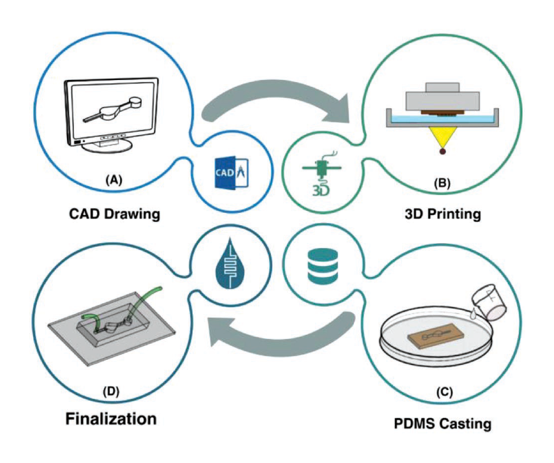
Methodology and Design
The fabrication process used in the study utilizes digital light processing (DLP) 3D printing. Device geometries are designed in CAD software (e.g., SolidWorks), then sliced and 3D printed. After printing, the post-processing workflow follows the mold undergoing: an isopropanol rinse → UV post-cure → PDMS casting → PDMS removal → plasma-bonding. During the PDMS casting stage, Sylgard 184 PDMS (10:1 base-to-curing-agent) was degassed for 15 minutes, poured directly onto the printed mold with no silanization or solvent treatment, oven-cured, peeled, punched, and plasma-bonded to glass or PDMS.
To benchmark the resin, four devices spanning both liquid-handling and biological applications were built and tested:
Spiral Separator
Pear-Shaped Microchamber
Hybrid Planar Micromixer
Tree-Shaped CGG
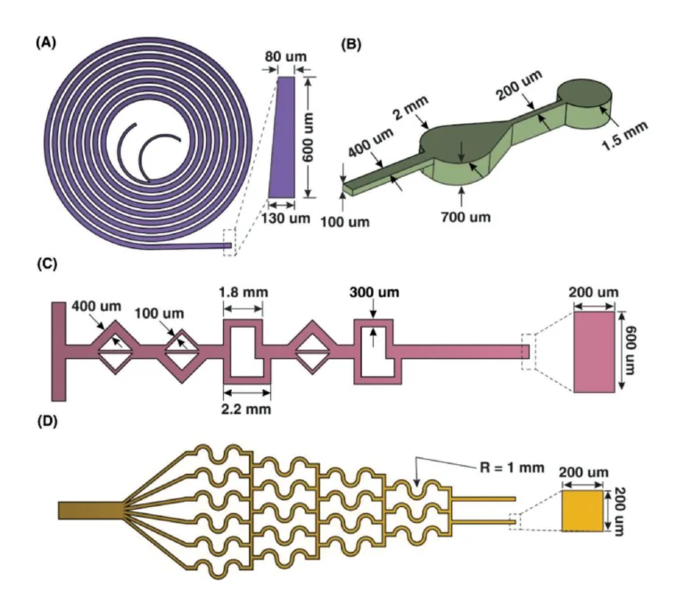
Figure 1. CAD schematics of the four benchmark geometries with channel dimensions — (A) spiral separator, (B) pear-shaped microchamber, (C) hybrid planar micromixer, (D) tree-shaped concentration gradient generator. Source: Bazaz et al. Rapid Softlithography Using 3D-Printed Molds. Adv Mater Technol. 2019;4(10).
The spiral microchannel is designed for particle separation applications. Its curved geometry enables inertial focusing, allowing particles to migrate to specific positions within the channel depending on flow rate. The design supports high flow rates, requiring strong bonding and smooth channel surfaces to maintain structural integrity and prevent leakage.
The hybrid micromixer integrates multiple mixing units to improve mixing efficiency under laminar flow conditions. In microchannels, where flow in predominantly laminar, mixing relies heavily on channel geometry rather than turbulence. The integration of different mixing structures enhances diffusion and fluid interaction.
For biological applications, the authors developed a pear-shaped microchamber designed to distribute fluid forces evenly and minimize shear stress during continuous perfusion and cell culture. This geometry supports cell viability by creating a more physiologically relevant microenvironment.
The final device is a tree-like concentration gradient generator splits and recombines two inlets across six outlets, producing a stable, stepped gradient for applications in drug-dosing studies.
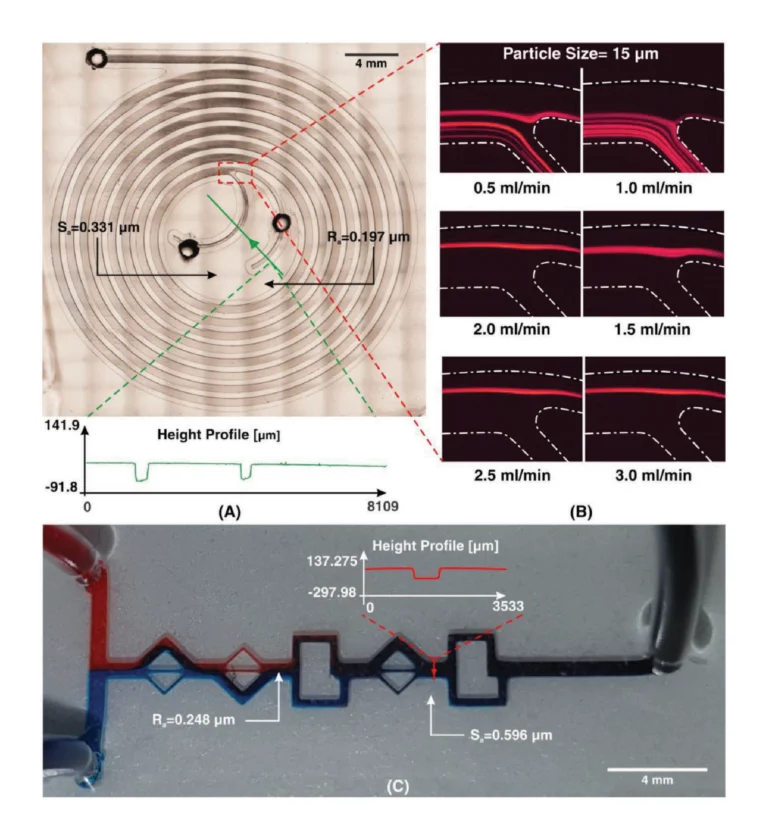
Figure 2. (A) Whole-chip bright-field image of spiral microchannel. (B) Experimental observation of 15 µm fluorescent beads at various flow rates from 0.5 to 3 mL min−1. C) Experimental observation of micromixer along the length of the microchannel. Source: Bazaz et al. Rapid Softlithography Using 3D-Printed Molds. Adv Mater Technol. 2019;4(10).
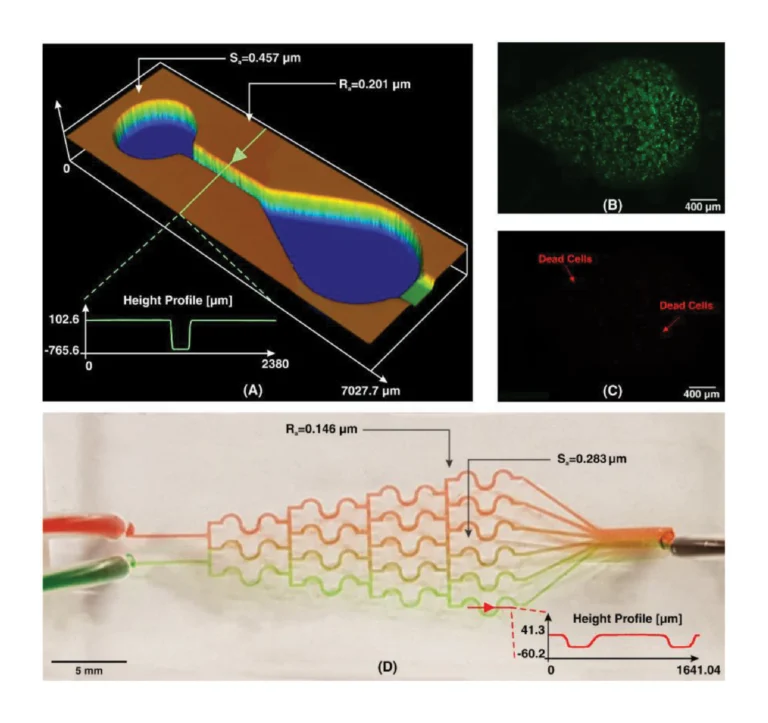
Figure 3. Whole-chip image of the cell culture device. B) Live and C) dead cell images after 24h incubation. D) Concentration gradient profile of two food colors (red and green). Source: Bazaz et al. Rapid Softlithography Using 3D-Printed Molds. Adv Mater Technol. 2019;4(10).
Results
Bazaz et al. (2019) investigate 3D printing as an alternative fabrication method to streamline the transition from design-to-device, and address key limitations of conventional lithographic approaches such as long fabrication times and complex workflows. As part of the study, the efficacy of a novel 3D printing resin was evaluated, an application-specific resin which enables the production of PDMS-compatible molds with minimal post-processing. To do so, the UTS research team 3D-printed and tested multiple devices architectures, including separators, mixers, cell culture platforms, and gradient generators.
Across all designs, surface quality and dimensional accuracy were critical performance metrics. Where conventional acrylate molds left an uncured PDMS film that causes replicas to stick and tear on demolding, PDMS cast onto the proposed resin detached cleanly and reproduced the mold pattern faithfully, with a smooth surface and no residual material on either part. Bonded devices showed no leakage across flow rates from 0.1 to 5 mL/min, and measured channel profiles matched the input CAD geometry.
The fabrication time for each device was significantly reduced to under five hours, with the cell-culture chamber mold printing in just 45 minutes. Additionally, the biocompatibility results were decisive. MCF-7 cells seeded into the PDMS microchamber and incubated for 24 hours at 37 °C showed more than 98% viability under live/dead staining, confirming that no cytotoxic residues were transferred from the printed mold to the cast PDMS replica. Because the resin requires no silanization, it eliminates a key step that typically makes standard 3D-printed molds unsuitable for cell-based applications.
As microfluidics continues to play a growing role in biomedical and analytical applications, this technique offers a scalable and accessible route to device fabrication, with the potential to accelerate innovation in the field.
“The present study can be considered as a milestone in the microfluidic field which can reduce the brainstorming-to-production from a time frame of several days … to less than 5 h.”
— Razavi Bazaz et al., Advanced Materials Technologies, 2019
Products Used In This Study

Questions on how our 3D printing system can work for your research? Connect with one of our specialists.
More Research Article Summaries
McGill University researchers build a 3D printed double-sided mold system to shape and merge brain organoids on a chip
University of Hawaiʻi researchers 3D print wearable "sweatainer" devices with true microscale internal channels
Want to read more article summaries?
We summarize the latest microfluidics and 3D printing research.
Curated from peer-reviewed publications featuring the CADworks3D system.