
Research Article Summary
5 Minute Read
University of Rhode Island Researchers 3D Print a Microfluidic Chip for Continuous Microplastics Detection
Title
A Low-Cost Microfluidic Method for Microplastics Identification: Towards Continuous Recognition
Authors
Pedro Mesquita, Liyuan Gong, Yang Lin
Journal
Micromachines, 2022, 13(4), 499
DOI Link
Summary
Mesquita et al. built a 3D-printed microfluidic chip that stains and flags microplastics in water on its own, replacing a slow, manual, batch-by-batch lab process with one continuous flow. The chip was then tested across several plastic shapes and fiber types to see how far its versatility would stretch.

Summary Author
This page was prepared by CADworks3D to summarize and highlight a peer-reviewed research article authored by independent researchers utilizing the CADworks3D system.
Title
A Low-Cost Microfluidic Method for Microplastics Identification: Towards Continuous Recognition
Authors
Pedro Mesquita, Liyuan Gong, Yang Lin
Journal
Micromachines, 2022, 13(4), 499
DOI Link
Key Results at a Glance
100X
Lowest Usable Dye Concentration
Diluting the Nile Red stain to 100X avoided particle clumping while still producing a readable signal
80°C
Best Staining Temperature
Fluorescence signal peaked at the hottest oven setting tested
10 Minutes
To Full Signal
Staining intensity leveled off after 10 minutes, marking the shortest reliable exposure window
6
Plastic and Fiber Types Validated
The same chip successfully stained microspheres, fragments, and both natural and synthetic fibers
Objective
Plastic pollution is a growing problem in oceans, rivers, and lakes, and most of it is not the large, visible kind. It is microplastics, plastic fragments smaller than 5 millimeters that come from broken-down packaging, synthetic fibers, and personal care products. Because they are small, light, and easy to mistake for other floating debris, microplastics are hard to track, and scientists still do not have a clear picture of how much plastic is out there, where it accumulates, or what it does to the organisms exposed to it.
Identifying microplastics is the first step toward answering those questions, but the tools currently used to do it come with tradeoffs. Techniques like Fourier transform infrared spectroscopy, Raman spectroscopy, and scanning electron microscopy can confirm exactly what a particle is made of, but they need expensive equipment and trained operators, which limits how much sampling can realistically get done. Visual inspection paired with fluorescent staining, usually with a dye called Nile Red, is cheaper and faster, but it is normally done one batch at a time in a lab. A researcher has to collect a water sample, bring it back, and stain it by hand. That works for a single snapshot, but it does not scale to long-term or continuous monitoring of a body of water.
Mesquita et al. set out to close that gap. Their goal was to combine Nile Red staining with microfluidics, a field built around moving tiny volumes of fluid through channels only a few millimeters wide, to see whether a small, low-cost, continuously running chip could do the same staining job as a manual lab process, without needing someone to babysit it.
Methodology and Design
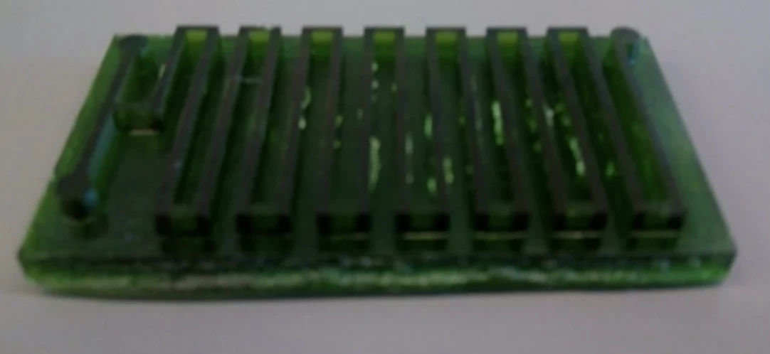
The chip started as a computer-aided design of a channel network with two inlets, one for the water sample carrying microplastics and one for the Nile Red dye, feeding into a long, winding serpentine channel that gives the two liquids room to mix before exiting through a single outlet. That design was 3D printed as a mold, and liquid PDMS (polydimethylsiloxane, a flexible silicone commonly used in lab-made microfluidic devices) was poured over it, cured, and peeled away to form the channel layer of the finished chip.

Figure 1. How the chip works: a water sample and Nile Red dye enter through separate inlets and mix inside a serpentine channel for continuous staining (left-A), compared to the manual, one-batch-at-a-time staining process (right-B). Source: Mesquita et al. A Low-Cost Microfluidic Method for Microplastics Identification: Towards Continuous Recognition. Micromachines. 2022.
With the chip built, the team ran it through a series of tests to confirm it could stain microplastics as reliably as a manual process, and to see how far its material versatility would stretch.
PE Microsphere Baseline
Multi-Plastic Panel
Fiber Comparison
Yeast Control
The first round of testing used polyethylene (PE) microspheres between 10 and 45 µm to work out the chip’s ideal operating conditions. The researchers varied Nile Red concentration, oven temperature, and residency time (how long the sample sat inside the channel), since all three affect how brightly a stained particle glows under fluorescence. Concentrations more dilute than 50X were needed to stop particles from clumping together within fractions of a second of contact, and once that threshold was set, 100X Nile Red at 80°C produced the strongest signal, reaching its peak after about 10 minutes.

Figure 2. The finished PDMS chip after bonding to a glass slide, ready for testing. Source: Mesquita et al. A Low-Cost Microfluidic Method for Microplastics Identification: Towards Continuous Recognition. Micromachines. 2022.
Once those settings were locked in, the same chip was challenged with a wider mix of materials: polystyrene (PS) microspheres, polypropylene (PP) and irregularly shaped PE fragments cut from plastic storage containers, and cotton and acrylic fibers pulled from clothing. PP and non-spherical PE produced the strongest fluorescence of any sample tested, and between the two fiber types, natural cotton stained more strongly than synthetic acrylic. Yeast was also run through the chip as a stand-in for natural organic matter, and it lit up almost as brightly as the plastics, a reminder that Nile Red staining alone cannot fully separate microplastics from organic particles and that a filtering step is still needed before deeper analysis. On the fabrication side, after the PDMS was cured in an oven overnight at 65°C, a corona treater was used to permanently bond the channel layer to a glass slide, and the finished chip was connected to a syringe pump so sample and dye could be pushed through continuously. With a channel 400 millimeters long and a 2 by 2 millimeter cross section, flow rates between 3.26 and 7.82 µL/min gave residency times of 5 to 12 minutes, letting the team dial in exactly how long each sample spent mixing with the dye.

Figure 3. The bonded chip running inside a lab oven, connected to inlet and outlet tubing and an external syringe pump for continuous flow. Source: Mesquita et al. A Low-Cost Microfluidic Method for Microplastics Identification: Towards Continuous Recognition. Micromachines. 2022.
Results
Across the static, manual staining tests, 100X Nile Red turned out to be the lowest concentration that avoided the clumping seen at higher concentrations, and it produced its strongest signal at 80°C, the hottest temperature tested. Staining intensity climbed steadily with time and flattened out at 10 minutes, meaning exposure beyond that point did not meaningfully improve the readout.
Moving those same settings into the microfluidic chip, slower flow rates, and therefore longer residency times, consistently produced brighter, more reliable staining, matching the trend seen in the manual tests. The chip’s best result, at a flow rate of 3.26 µL/min, still measured roughly 37% dimmer than the equivalent manual sample, likely because gentle mixing by diffusion inside the channel is less thorough than actively shaking a tube by hand. Even so, the chip stained particles continuously and without any manual pipetting, which the manual process cannot offer.
Material testing confirmed the chip works well beyond a single type of plastic. It successfully stained and identified 6 distinct plastic and fiber types, including PE and PS microspheres, PP and non-spherical PE fragments, and both cotton and acrylic fibers. Non-spherical fragments produced the highest fluorescence overall, and natural cotton outperformed synthetic acrylic. The one limitation the team flagged was organic interference, since yeast particles fluoresced at levels comparable to the plastics themselves.
“Low-cost fabrication methods such as 3D printing and molding can be applied to further minimize the cost associated with this method.”
— Mesquita et al., Micromachines (2022)
Products Used In This Study
Questions on how our 3D printing system can work for your research? Connect with one of our specialists.
More Research Article Summaries
McGill University researchers build a 3D printed double-sided mold system to shape and merge brain organoids on a chip
University of Hawaiʻi researchers 3D print wearable "sweatainer" devices with true microscale internal channels
Want to read more article summaries?
We summarize the latest microfluidics and 3D printing research.
Curated from peer-reviewed publications featuring the CADworks3D system.