
Research Article Summary
5 Minute Read
University of Toronto researchers build a microfluidic chip that sorts tumor cells by surface signature
Title
Tracking the dynamics of circulating tumour cell phenotypes using nanoparticle-mediated magnetic ranking
Authors
Mahla Poudineh, Peter M. Aldridge, Sharif Ahmed, et al.
Journal
Nature Nanotechnology, (2016)
DOI Link
Summary
Poudineh et al. developed a nanoparticle-based microfluidic chip that sorts individual circulating tumour cells (CTCs) into one of 100 capture zones based on how strongly each cell expresses a chosen surface marker, turning a rare-cell detection problem into a single-cell resolution phenotyping tool that works directly on unprocessed whole blood.

Summary Author
This page was prepared by CADworks3D to summarize and highlight a peer-reviewed research article authored by independent researchers utilizing the CADworks3D system.
Title
Tracking the dynamics of circulating tumour cell phenotypes using nanoparticle-mediated magnetic ranking
Authors
Mahla Poudineh, Peter M. Aldridge, Sharif Ahmed, et al.
Journal
Nature Nanotechnology, (2016)
DOI Link
Key Results at a Glance
100
Capture Zones
Each zone ranks cells by exact surface marker density, not just presence or absence
10 cells/mL
Detection Sensitivity
Tumor cells were identified even at whole-blood concentrations near the noise floor of older methods
> 90%
Recovery Efficiency
Four cancer cell lines with very different marker levels were all captured at similarly high rates
99.98%
White Blood Cell Depletion
Nearly all normal blood cells were excluded, leaving a clean population of tumor cells to analyze
Objective
When a solid tumor starts to spread, it sheds cells into the bloodstream. These circulating tumour cells (CTCs) are a live snapshot of a cancer’s biology, and in principle they could tell doctors which tumors are becoming more aggressive long before a scan would show it. In practice, CTCs are extraordinarily hard to study. They show up at roughly one cell for every billion normal blood cells, and the cells that are present are not identical to one another. A single tumor can shed cells carrying very different combinations of surface proteins, and those combinations shift over time as cells move from a stationary, epithelial state toward a more mobile, invasive one.
Flow cytometry, the standard tool for sorting cells by surface marker, typically needs tens of thousands of cells for a reliable readout, far more than a CTC blood sample can supply. Microfluidic chips built for CTCs solve the rarity problem: they can catch and count these cells from a full blood sample. What most cannot do is say anything about the cells beyond the fact that they were caught. Pulling a cell out of the bloodstream is not the same as reporting how much of a given surface marker that cell was carrying.
The authors set out to close that gap with a device sensitive enough to work directly on unprocessed whole blood, precise enough to sort CTCs by degree of marker expression rather than a simple present or absent call, and detailed enough to track how that expression changes as a tumor grows and becomes more aggressive.
Methodology and Design
The device, called Magnetic Ranking Cytometry (MagRC), is a microfluidic chip patterned with a graded array of nickel micromagnets seated inside X-shaped channel structures. The chip is built in layers: a nickel film is patterned into circular micromagnets that grow larger along the length of the channel, a photoresist layer defines the X-shaped channel geometry above the magnets and serves as the mold for the channel network, and a molded PDMS layer is bonded on top to seal everything into a finished chip. Because the micromagnets increase in size from inlet to outlet, the local magnetic field gradient, and therefore the pull on a magnetically tagged cell, increases predictably along the chip, creating 100 distinct capture zones.
This graded architecture let the researchers apply the same chip across four progressively more demanding test scenarios, moving from cultured cells to real clinical blood samples.
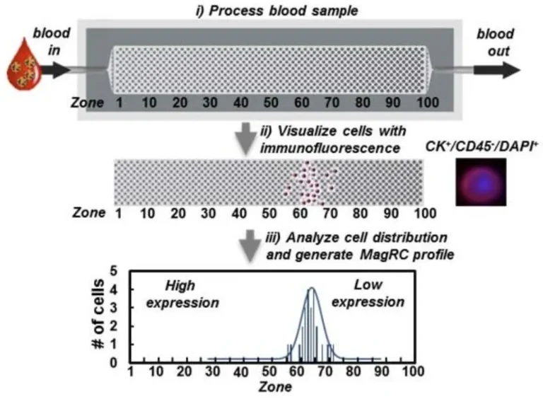
Figure 1.
Cell Line Panel
Spiked Whole Blood
Xenograft Mouse Model
Patient Blood Samples
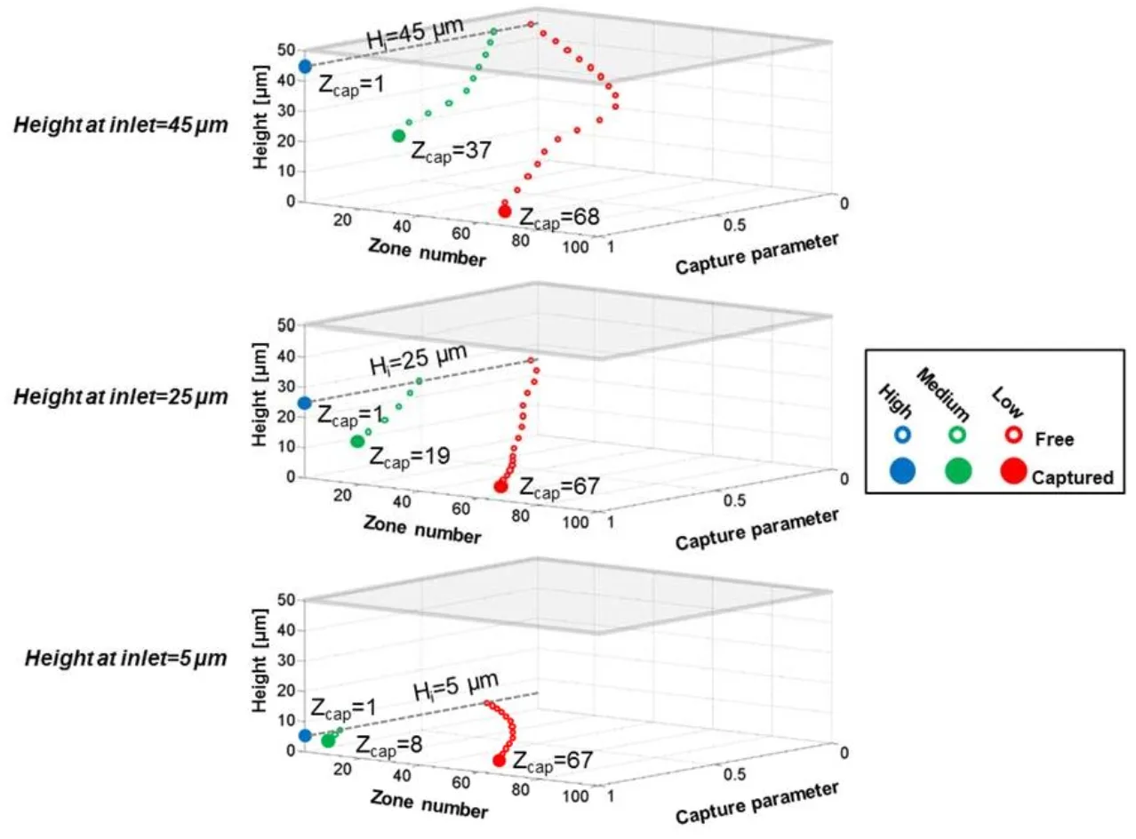
Blood or buffer samples are first incubated with magnetic nanoparticles coated in an antibody against a chosen surface marker, most often EpCAM, a protein many tumor cells lose as they become more invasive. The sample is then pumped through the chip, where each cell settles into the capture zone matching its magnetic loading: heavily tagged, high-expression cells stop early in the chip where the micromagnets are small, while lightly tagged, low-expression cells travel further before the larger downstream magnets are able to hold them.
Four breast and prostate cancer cell lines with known, very different EpCAM levels were run through the chip first to confirm that each line produced a distinct, reproducible capture profile and that even low-expression cells were recovered at high rates. The chip was then challenged with unprocessed whole blood spiked with as few as 10 tumor cells per milliliter, tested side by side against the clinical gold standard CellSearch system and against flow cytometry, and it held its sensitivity in this messier sample type where the comparison methods lost significant ground. From there, the chip was used to draw blood every 10 days from mice carrying human breast tumors, tracking how the captured CTC profile shifted position as the tumors grew and, in one group, became metastatic. Finally, the same chip profiled blood from prostate cancer patients, both localized and metastatic cases, to see whether tumor grade tracked with any pattern in CTC surface marker expression.
On the fabrication side, the nickel layer is patterned using standard contact lithography and wet etching, and the channel mold is built from a 50 µm layer of SU-8 photoresist that is soft-baked, exposed, and developed on top of the patterned nickel. A PDMS layer is then cast over this mold, punched for inlet and outlet ports, and sealed onto the substrate to complete the channel. Antibody-coated magnetic nanoparticles were incubated with each sample for 30 minutes before it was pumped through the chip at a controlled flow rate, followed by a buffer wash and on-chip immunostaining to distinguish tumor cells from any white blood cells that bound non-specifically.
Results
Across the four cell lines tested, MagRC produced clearly separated capture profiles that lined up with each line’s known EpCAM level, spreading cells across the chip’s full span of 100 capture zones rather than sorting them into a single yes or no gate. Recovery efficiency stayed above 90% for every line tested, including the two with the lowest marker expression, which are exactly the phenotypes older marker-dependent methods tend to miss.
That sensitivity carried over into unprocessed whole blood. MagRC reliably profiled samples spiked with as few as 10 tumor cells per milliliter. In head-to-head testing, it recovered low-EpCAM cells at far higher rates than the CellSearch system, which is tuned mainly for high-EpCAM cells, and it produced a usable signal in whole blood where flow cytometry could not; flow cytometry only worked after red blood cells were lysed, a step that discards over half the tumor cells before they can even be counted. Purity was high throughout, with the chip excluding 99.98% of white blood cells.
In the mouse model, CTC counts rose as tumors grew in both study groups, but only the faster-growing, estrogen-driven tumors produced a clear phenotypic shift toward lower EpCAM expression, alongside the appearance of lung micrometastases. In patient samples, CTCs from metastatic prostate cancer clustered consistently in the low-EpCAM zones, while CTCs from localized cases spread more widely and in a pattern that tracked with tumor grade, from earlier zones in low-grade tumors to later zones in high-grade ones.
“A chip that can only tell you a cell is there isn’t enough. Real insight into a tumor’s behavior comes from knowing how much of a marker each single cell carries, and how that changes over time.”
— Poudineh et al., Nature Nanotechnology, 2016
Products Used In This Study

Questions on how our 3D printing system can work for your research? Connect with one of our specialists.
More Research Article Summaries
McGill University researchers build a 3D printed double-sided mold system to shape and merge brain organoids on a chip
University of Hawaiʻi researchers 3D print wearable "sweatainer" devices with true microscale internal channels
Want to read more article summaries?
We summarize the latest microfluidics and 3D printing research.
Curated from peer-reviewed publications featuring the CADworks3D system.