
Research Article Summary
5 Minute Read
NC State and UNC researchers build a zero-power microneedle patch for painless interstitial fluid sampling using 3D printed molds
Title
Design and characterization of a self-powered microneedle microfluidic system for interstitial fluid sampling
Authors
Christopher T. Sharkey, Angélica F. Aroche, Isabella G. Agusta, et al.
Journal
Lab on a Chip, 2025, Volume 25, Pages 4577–4587
DOI Link
Summary
Researchers developed a fully passive, wearable microneedle device that pulls interstitial fluid from skin models and recovers the stress hormone cortisol without any electrical power, combining hydrogel-forming microneedles, a paper microfluidic channel, and an osmotic pump. Several rigid housing components were cast from 3D printed molds, enabling rapid, low-cost fabrication of the device assembly (Sharkey et al.).
Summary Author
This page was prepared by CADworks3D to summarize and highlight a peer-reviewed research article authored by independent researchers utilizing the CADworks3D system.
Title
Design and characterization of a self-powered microneedle microfluidic system for interstitial fluid sampling
Authors
Christopher T. Sharkey, Angélica F. Aroche, Isabella G. Agusta, et al.
Journal
Lab on a Chip, 2025, Volume 25, Pages 4577–4587
DOI Link
Key Results at a Glance
50 µm
Mold Layer Height
Print resolution used to fabricate the negative molds for the casting device housing
~2 µm
Parylene C coating
Thin release layer applied to printed molds so cured silicone lifts out cleanly
< 10 min
Full-Channel Flow
Fluid and dye traveled the entire paper microfluidic channel in under ten minutes
15 min
Rapid Cortisol Recovery
Accurate cortisol quantification achieved in as little as a 15-minute application
Objective
Interstitial fluid (ISF) is the watery fluid that sits in the spaces between cells, and it is one of the most promising fluids for at-home and point-of-care health monitoring. Unlike sweat, which mostly carries small molecules like glucose and lactate, ISF closely mirrors blood. Prior work has shown that 90 to 99 percent of blood proteins also appear in ISF, and some proteins show up in ISF that are not found in blood at all. That makes ISF a rich, fast-responding window into what is happening inside the body, with very little delay compared to blood.
The problem is access. ISF is most abundant in the dermis, sitting under the stratum corneum and the viable epidermis, which together form a barrier roughly 50 to 200 µm thick. Conventional ways of pulling ISF out, such as suction blistering, microdialysis, and open-flow microperfusion, are painful, slow, and invasive, which makes them poorly suited for everyday use. For a wearable device to be practical, it needs to reach ISF gently, collect enough of it (roughly 1 to 20 µL), and do all of this without external power or bulky equipment.
Microneedles are a strong candidate for this job. Measuring a few hundred µm to about a millimeter long, they pierce the outer skin layers while staying above the nerve-rich and blood-rich deeper dermis, so insertion is nearly painless and draws minimal blood. Hydrogel-forming microneedles are especially appealing because they swell and soak up fluid on contact, are biocompatible, and are inexpensive to make. Their weakness is that they tend to collect smaller fluid volumes on their own.
This paper addresses a specific gap. Many earlier integrated systems relied on active pumping or complex multilayer architectures, which are hard to scale and hard to translate into real products. The authors set out to design a wearable system that is genuinely passive, simple, and cheap to build, while still extracting ISF reliably and recovering a clinically meaningful biomarker. Achieving that low-cost, simple-to-build goal depended heavily on how the device housing was fabricated, which is where rapid molding from 3D printed parts comes in.
Methodology and Design
The fabrication process behind this device centers on stereolithography (SLA) 3D printing. Components are designed as negative molds in CAD software, then 3D printed using a clear microfluidic resin. The post-processing workflow follows the mold undergoing: an isopropyl alcohol rinse → UV post-cure → parylene C coating → PDMS casting → cure → demold.

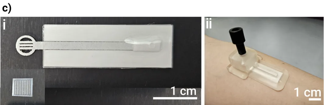
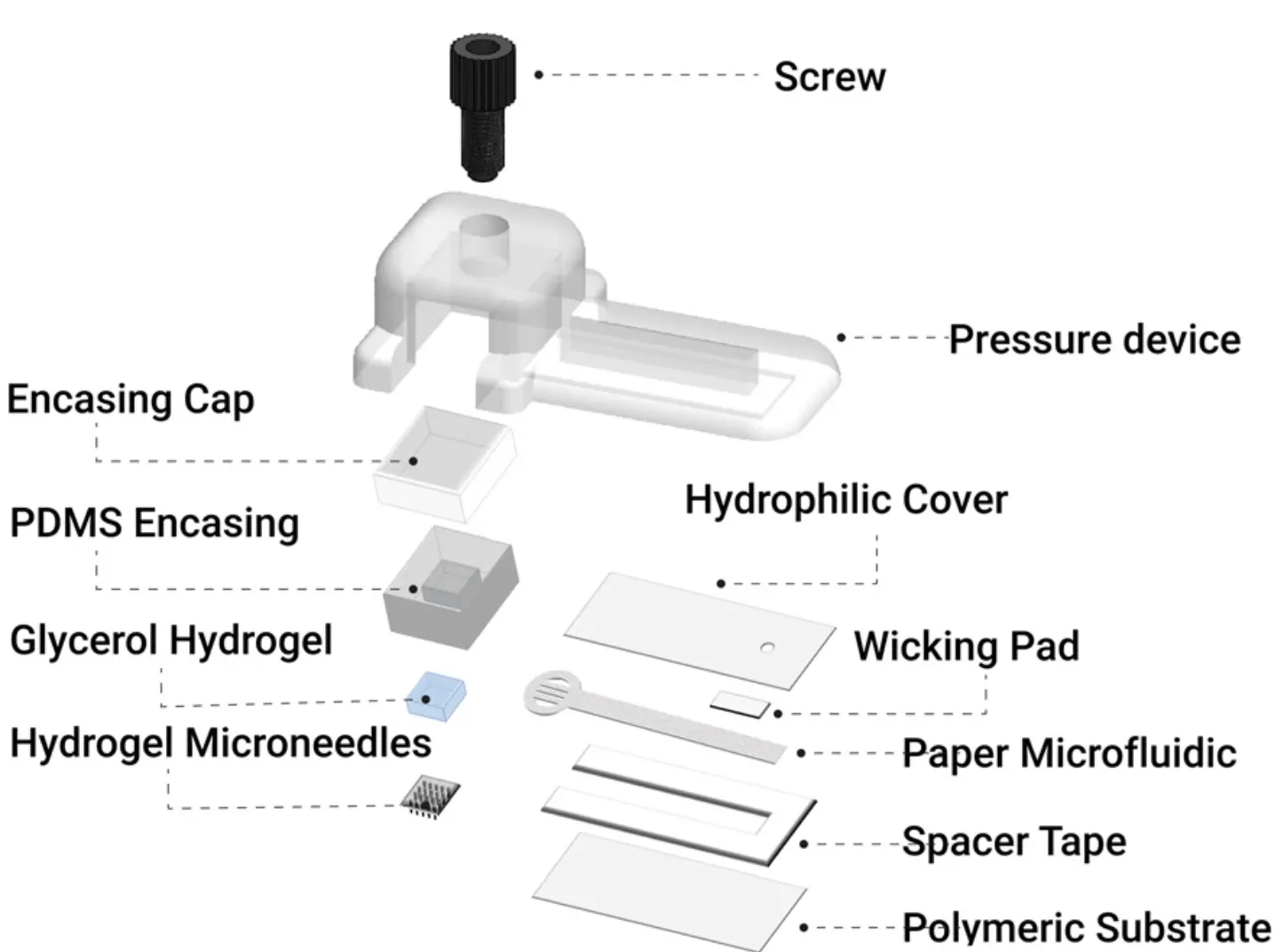
Figure 1. A schematic of the patch on skin showing how fluid moves through it, a labeled exploded view of every component, and photographs of the assembled microneedle patch and paper microfluidic applied to a forearm. Source: Sharkey et al. Design and characterization of a self-powered microneedle microfluidic system for interstitial fluid sampling. Lab on a Chip. 2025.
The device brings together four functional pieces that each do one job, and the way they were manufactured is central to making the whole system fast and affordable to produce.
MeHA Microneedle Array
Paper Microfluidic Channel
Glycerol Osmotic Pump
Rigid Housing and Applicator
The microneedles were made from methacrylated hyaluronic acid (MeHA). A liquid MeHA and photoinitiator solution was drop-cast into PDMS (silicone) molds shaped as a 10 by 10 array of pyramidal needles, each 800 µm tall. The filled molds were centrifuged to drive the solution into the needle tips, dried, removed, and then UV crosslinked to lock in their shape. On contact with fluid, these needles swell and absorb ISF.
The osmotic pump is the part that generates suction without any power. Polyacrylamide hydrogels were soaked in concentrated glycerol, which creates a strong osmotic pressure gradient that passively draws fluid up through the device. Glucose was also tested as an alternative driving agent. To carry that fluid sideways for analysis, the team built a paper microfluidic channel from chromatography paper, cut to shape with a laser cutter, then layered with a transport film, spacer tape, and a polymer backing so the channel would not deform.
The rigid housing and applicator relied on the same 3D printing pipeline described above. The silicone (PDMS) casing that holds the osmotic pump, along with a rigid cap and an external pressure device, all started as negative molds designed in CAD. Those molds were 3D printed on a stereolithography printer using a clear microfluidic resin at a 50 µm layer height. After printing, the molds were rinsed in isopropyl alcohol, UV cured, and coated with about 2 µm of parylene C so the cured silicone would release cleanly. Silicone was then mixed, degassed, cast into the printed molds, degassed again, and cured.
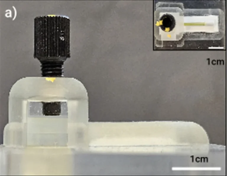
Figure 2. Side and top-down views of the complete device in use, showing the 3D printed rigid housing, the screw-based pressure device that applies steady downward force, and the microneedle patch pressed against the skin substitute. Source: Sharkey et al. Design and characterization of a self-powered microneedle microfluidic system for interstitial fluid sampling. Lab on a Chip. 2025.
To test the assembled system, the researchers used two skin substitutes, an agarose gel and a commercial synthetic skin, each wrapped in stretched Parafilm to mimic the resistance of real skin. They tracked how fast fluid and dye moved through the paper channel, compared the osmotic pump against a solid silicone block as a control, and measured cortisol recovery using ELISA across loaded concentrations of 0, 12, 48, and 96 ng/mL over 15-minute, 45-minute, and 24-hour windows.
Results
The microneedles performed as intended. Electron microscopy confirmed clean, well-formed needles, and penetration tests showed they consistently pierced the skin models. Within just 30 seconds of fluid contact, individual needles swelled by an average of 72% in deionized water and 58% in PBS, demonstrating rapid uptake.

Figure 3. A close-up scanning electron micrograph of the pyramidal needle array, showing the clean, well-formed needles produced by casting methacrylated hyaluronic acid in the molds. Source: Sharkey et al. Design and characterization of a self-powered microneedle microfluidic system for interstitial fluid sampling. Lab on a Chip. 2025.
Fluid transport through the paper channel was both fast and dependable. Across every combination of skin model and pump type, fluid and dye reached the full length of the paper channel within 10 minutes. In agarose models, fluid flowed at roughly 4 to 5.5 µL/min, while in synthetic skin the range was 2 to 5 µL/min. The solid silicone control actually pushed slightly higher average flow rates than the osmotic pump, but the osmotic pump produced lower standard deviations, meaning more predictable and uniform extraction. That consistency matters more than raw speed for reliable diagnostics, and the authors expect the osmotic approach to perform even better on real skin, where mechanical squeezing is less reliable.
Cortisol recovery was the headline biological result. Without the paper microfluidics, recovered cortisol tracked the starting skin concentration almost perfectly, with R² values above 0.99 at both 45 minutes and 24 hours. Interestingly, the needles recovered two to three times more cortisol at 45 minutes than at 24 hours, pointing to depletion or degradation over long wear. With the paper microfluidics integrated, the strong linear relationship held at 15 and 45 minutes, with R² values above 0.9, confirming that accurate cortisol quantification is achievable in as little as 15 minutes.
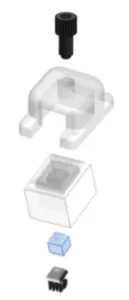
Figure 4. A CAD-style exploded view of the molded housing and osmotic pump used in the baseline cortisol experiments, run without the paper microfluidics.Source: Sharkey et al. Design and characterization of a self-powered microneedle microfluidic system for interstitial fluid sampling. Lab on a Chip. 2025.
The spatial analysis offered a useful design lesson. After 15 minutes, cortisol levels in the paper segments farthest from the needles had not fully equilibrated with the skin concentration, an effect that mostly resolved by 45 minutes. The practical takeaway is that biosensors should be placed close to the extraction site for short-duration sampling. Over 24 hours, recovery from the paper dropped at lower concentrations, likely from cortisol instability and evaporation, though the device still cleanly distinguished the highest concentration of 96 ng/mL. Finally, swapping glycerol for glucose as the osmotic driver produced negligible differences in cortisol recovery, showing the system tolerates flexible material choices, an advantage when integrating future biosensors.
“A negative mold was designed in SolidWorks 2023 and printed on a CADWorks3D Profluidics 285D stereolithography 3D printer using Clear Microfluidic Resin with a layer height setting of 50 μm.“
— Sharkey et al., Lab on a Chip, 2025
Products Used In This Study
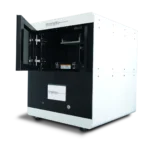
Questions on how our 3D printing system can work for your research? Connect with one of our specialists.
More Research Article Summaries
McGill University researchers build a 3D printed double-sided mold system to shape and merge brain organoids on a chip
University of Hawaiʻi researchers 3D print wearable "sweatainer" devices with true microscale internal channels
Want to read more article summaries?
We summarize the latest microfluidics and 3D printing research.
Curated from peer-reviewed publications featuring the CADworks3D system.